Anatomy and Physiology
Illustrations
Copyright © July 2004 Ted Nissen
TABLE OF
CONTENTS
8 Skeletal System-Appendicular
Skeleton
13 Spinal Cord and Spinal Nerves
14 The Brain and Cranial Nerves
15 Sensory, Motor, and Integrative
Systems
19 Cardiovascular System-Blood
20 Cardiovascular System-Heart
21 Cardiovascular System-Vessels
and Routes
22 Lymphatic and Immune System
27 Fluid, Electrolyte, and
Acid-Base Dynamics
29 Development and Inheritance
1 Introduction
Back
Table of Contents References
1.1 Planes of Reference
Back
Table of Contents References
2 Chemical Organization
Back
Table of Contents References
3 Cellular Organization
Back
Table of Contents References
4 Tissue Organization
Back
Table of Contents References
4.1 Tendon, Ligament, Muscle Tissue and Cells
Back
Table of Contents References
4.2 Synovial (Mucous) Sheaths of the Tendons & Retinaculum (Ligaments) Around the Ankle (Lateral & Medial)
Back
Table of Contents References
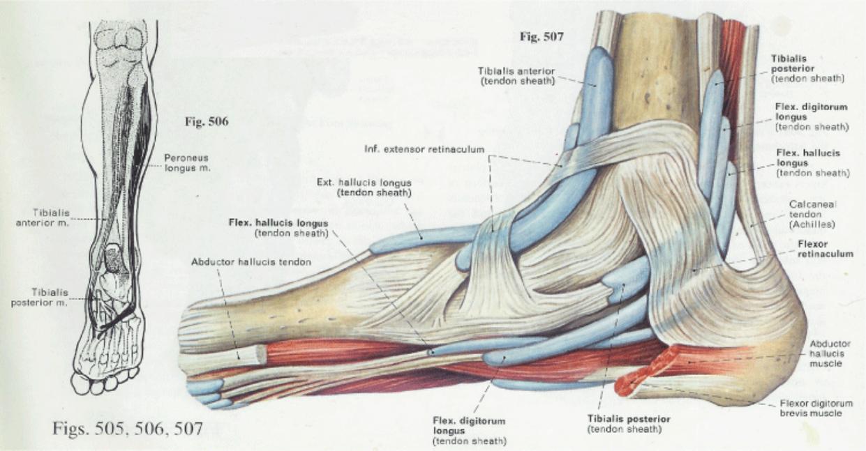
4.3 Fig. 505 Tendons at the Ankle Region Dorsolateral View (Right Foot)
4.3.1 Similar to what is observed at the wrist, tendons at the ankle region passing from the leg into the foot are bound by closely investing retinacula. The tendons themselves are surrounded by synovial sheaths, which are indicated in blue in this figure and in Fig. 507.
4.3.2 Anterior to the ankle joint and on the dorsum of the foot are three separate synovial sheaths. One is for the extensor digitorum longus and peroneus tertius, a second is for the extensor hallucis longus and the third surrounds the tibialis anterior (see Fig. 507). Behind the lateral malleolus is a single tendon sheath for the peroneus longus and brevis, which then splits distally to continue along each individual tendon for some distance. The inferior extensor retinaculum and the superior and inferior peroneal retinacula which bind the tendons and their sheaths close to bone.
4.3.3 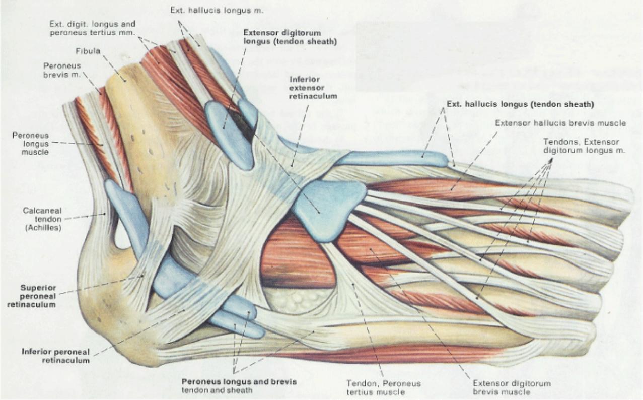
4.4 Fig 506 The Tendons of the Peroneus Longus and Tibialis Anterior Muscle
4.4.1 The tendons of the tibialis anterior and peroneus longus muscles insert on the medial aspect of the plantar surface of the foot. The peroneus longus muscle achieves this insertion by traversing the sole of the foot from lateral to medial. In this manner, the two muscles form a tendinous sling under the foot, which serves to support the transverse arch. Also assisting in this support is the tendon of the tibialis posterior muscle.
4.4.2 Fig 507 Tendons at the Ankle Region Medial View (Right Foot)
4.4.2.1 From this medial view can be seen the synovial sheaths and tendons of the tibialis anterior and extensor hallucis longus on the dorsum of the foot, as well as the three tendons which course beneath the medial malleolus from the posterior compartment of the leg into the plantar foot: tibialis Posterior. flexor digitorum longus and flexor hallucis longus.
4.4.2.2 The bifurcating nature of the inferior extensor retinaculum, and the manner in which the flexor retinaculum secures the structures beneath the medial malleolus
4.4.2.3
5 Integumentary System
Back Table of Contents References
6 Skeletal Tissue
Back
Table of Contents References
7 Skeletal System-Axial
Back
Table of Contents References
8 Skeletal System-Appendicular Skeleton
Back
Table of Contents References
8.1 Fig. 480 The Tibiofibular Union and Interosseous Membrane (Right Leg)
8.1.1 From this anterior view that the shafts of the fibula and the tibia are connected from the knee to the ankle by the interosseous membrane. Additionally, the two bones are joined proximally (the tibiofibular joint) and distally (the tibiofibular syndesmosis).
8.1.2 Proximally, the head of the fibula articulates with the inferolateral aspect of the lateral condyle of the tibia. This is a gliding joint, surrounded by an articular capsule and strengthened by the anterior and posterior ligaments of the head of the fibula.
8.1.3 The ligamentous union between the distal ends of the fibula and tibia is formed by the anterior and posterior tibiofibular ligaments
8.1.4
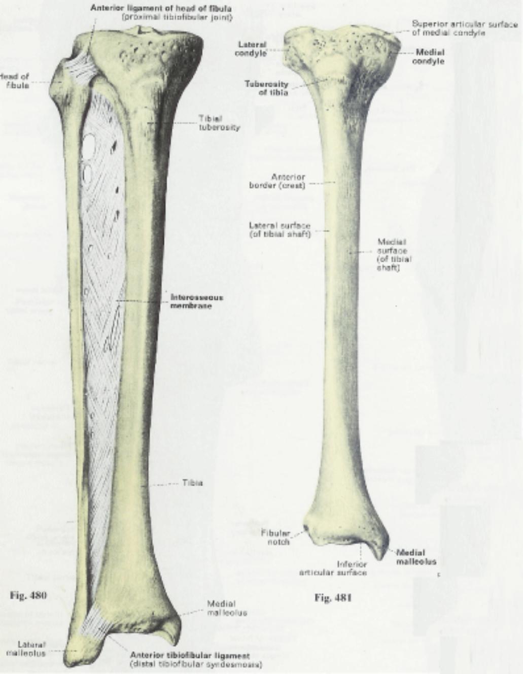
8.1.5 Fig. 481 The Right Tibia Anterior View
8.1.5.1 Note that the proximal extremity is marked by the two tibial condyles and the tibial tuberosity. The medial aspect of the distal extremity forms the medial malleolus.
8.2 Fig. 484 The Right Tibia Posterior View
8.2.1 The smooth posterior surface of the shaft of the tibia is marked by a prominent ridge (the soleal line) and a large oblong foramen (the nutrient foramen). The tibial shaft tapers toward a larger proximal extremity and somewhat less pronounced distal extremity.
8.2.2 At the proximal extremity the rounded medial and lateral condyles are separated by the intercondylar eminence, anterior and posterior to which attach the cruciate ligaments. At its distal extremity, the tibia articulates with the talus and, on this posterior surface, presents grooves for the passage of the tendons of the tibialis posterior, flexor digitorum longus and flexor hallucis longus.
8.2.3
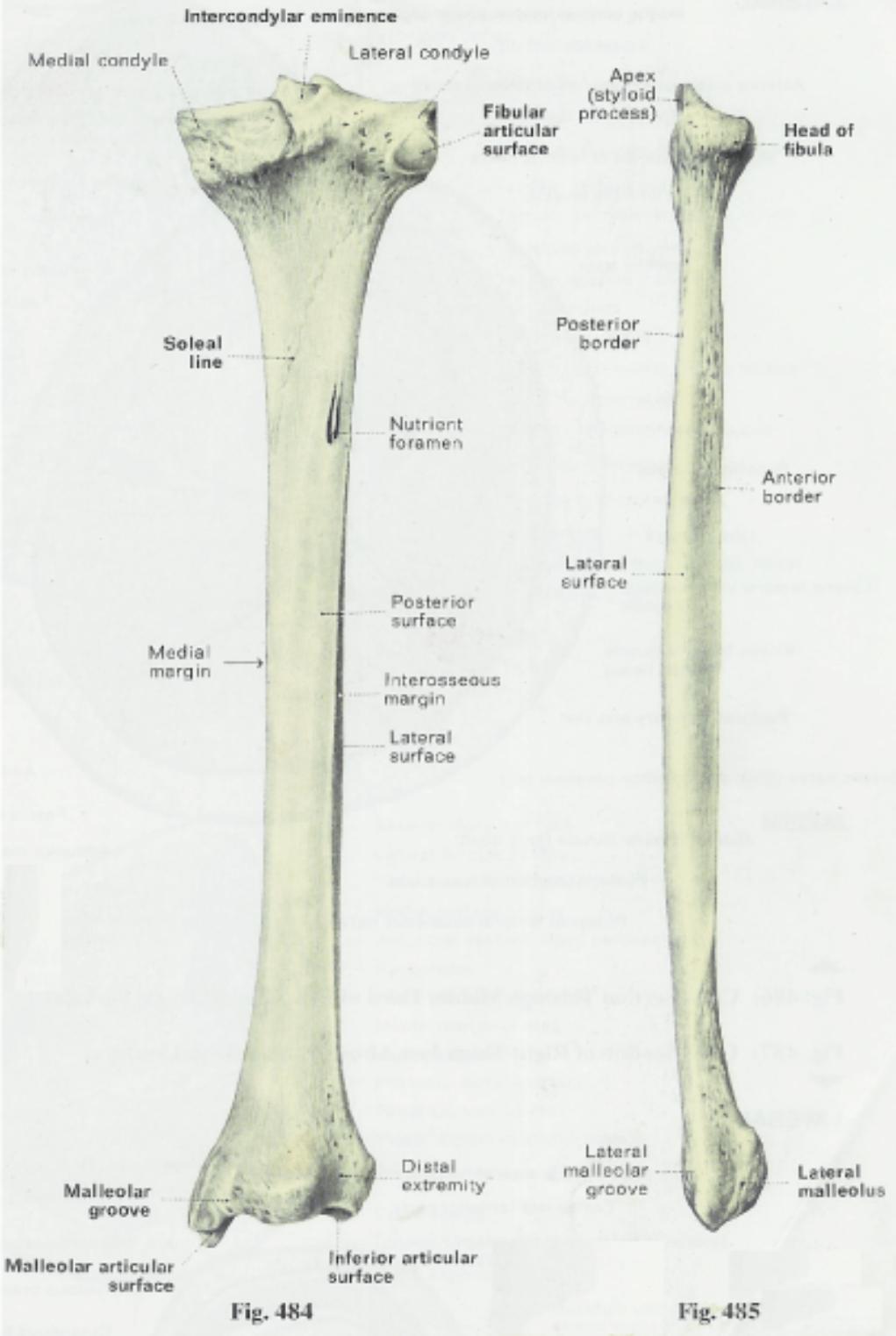
8.2.4 Fig 485 The Right Fibula Lateral View
8.2.4.1 The fibula is a long slender bone situated lateral to the tibia to which it articulates proximally (see Figure 480). Distally, the fibula expands to form the lateral malleolus. The medial aspect of its inferior articular surface participates with the tibia in forming the talocrural or ankle joint.
8.2.4.2 Although the fibula does not bear any weight of the trunk (since it does not participate in the knee joint articulation), it is important because of the numerous muscles which attach to its surface (see Figures 482 and 483) and because it assists in the formation of the ankle joint.
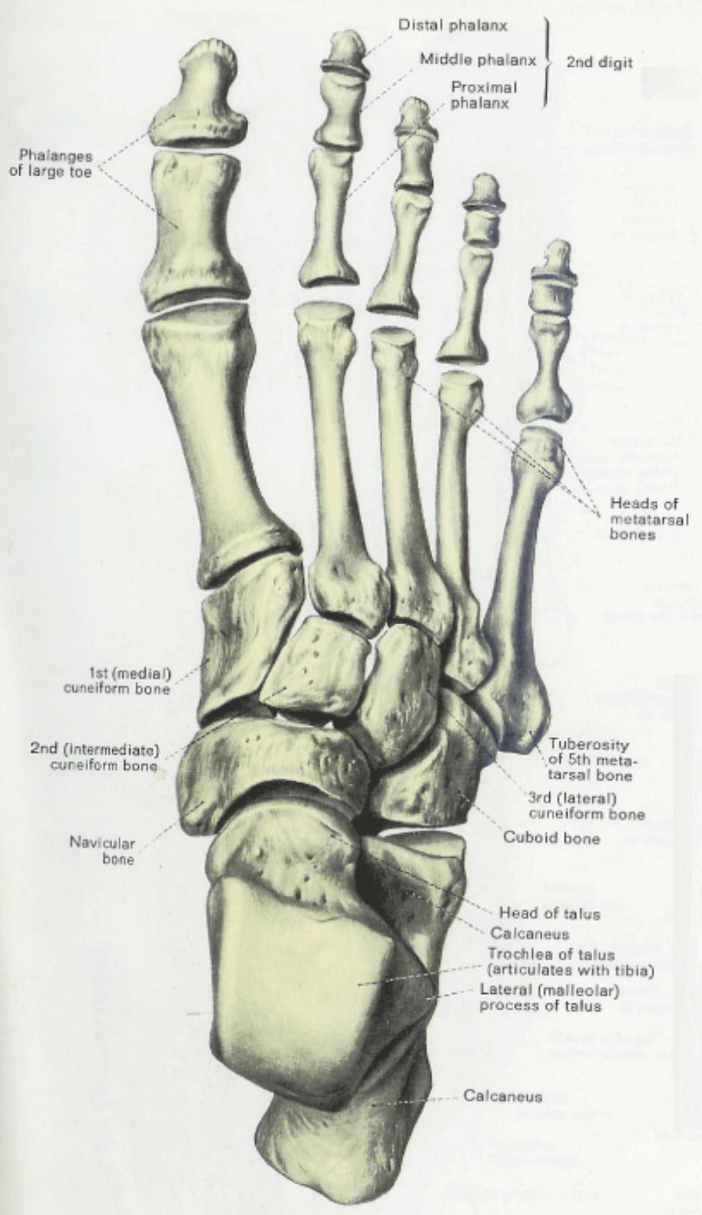
8.3 Fig 501 The Bones of the right Foot, Dorsal View
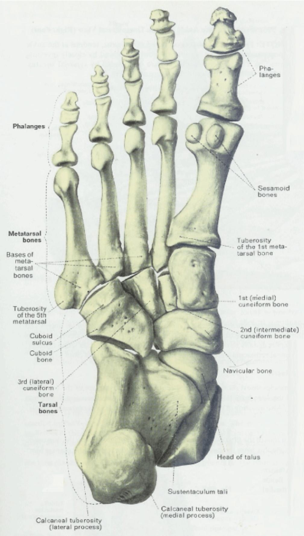
8.3.1 The skeleton of the foot consists of 7 tarsal bones, 5 metatarsal bones and 14 phalanges. The toes are numbered in order from medial to lateral so that the large toe is the 1st digit while the small toe is the 5th digit.
8.3.2 The weight of the body is transmitted by the tibia to the talus, which then redistributes this weight to the calcaneus inferiorly (the "heel" of the foot) and the navicular bone distally (toward the heads of the metatarsals and the "ball" of the foot)
8.3.3 Distal to the navicular and calcaneus are the three cuneiform bones and the cuboid; these then articulate with the individual metatarsal bones of the digits. Observe the similarity of the anatomy of the skeleton of the human foot and the hand, but appreciate their marked differences in function.
8.3.4
8.4 Fig 503 The Bones of the Right Foot Plantar View
8.4.1 The largest bone in the foot is the calcaneus. From this surface can be seen the prominent calcaneal tuberosity which projects posteriorly and inferiorly (forming the heel) and the sustentaculum tali, the dorsal surface of which contains articular facets for the talus.
8.4.2 The cuboid bone and the sulcus on its plantar surface for the passage of the peroneus longus tendon across the sole of the foot.
8.4.3 The long, slender metatarsal bones which are curved, such as to be concave on their plantar surface and convex dorsally. Observe the large tuberosity on the lateral side of the base of the 5th metatarsal.
8.4.4
8.5 Right Foot Dorsal/Plantar
Back
Table of Contents References
8.6 Left Talus
Back Table of Contents References
8.7 Calcaneus
Back
Table of Contents References
8.8 Navicular and Cuboid Bones
Back
Table of Contents References
8.9 1st, 2nd, and 3rd Cuneiform Bones
Back
Table of Contents References
8.10 Metatarsals
Back
Table of Contents References
9 Articulations
Back Table of Contents References
9.1 Right Foot Lateral & Medial Ligaments
Back
Table of Contents References
9.2 Plantar Ligaments & Joints
Back
Table of Contents References
9.3 Right Foot Synovial Joint Cavities
Back
Table of Contents References
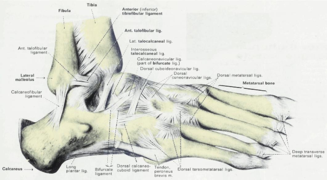
9.4 Fig 508 Ligaments of the Ankle and Foot Dorsolateral View (Right Foot)
9.4.1 The fibula is attached to the tibia distally by the anterior (inferior) tibiofibular ligament. Additionally, the lateral malleolus of the fibula is attached to the talus by the relatively weak anterior and posterior (Fig. 512) talofibular ligaments, and to the calcaneus by the calcaneofibular ligament
9.4.2 The joint between the talus and calcaneus (subtalar joint) is principally strengthened by the interosseous talocalcaneal ligament. The talocalcaneonavicular joint more anteriorly is of important clinical significance since the weight of the body tends to push the head of the talus down between the navicular and calcaneus. The stability of this joint is assisted dorsolaterally by the calcaneonavicular ligament (a part of the bifurcate ligament); however, the thick plantar calcaneonavicular or spring ligament (Figs. 509, 511,513,514) is the principal support for this joint in the maintenance of the longitudinal arch of the foot.
9.4.3 The bifurcate ligament consists of the calcaneonavicular ligament and the calcaneocuboid ligament.
9.4.4
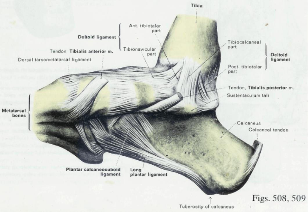
9.5 Fig 509 Ligaments of the Ankle and Foot Medial View (Right Foot)
9.5.1 The medial aspect of the ankle joint is protected by the deltoid ligament, which is triangular in shape and which connects the tibia (medial malleolus) to the navicular, calcaneus and talus. The deltoid ligament consists of 4 parts: a) an anterior part which attaches the medial malleolus to the navicular (tibionavicular part), b) a superficial part attaching the malleolus to the sustentaculum tali ofthe calcaneus (tibiocalcaneal part), and c) and d) the anterior and posterior tibiotalar parts which lie more deeply and attach the malleolus to the adjacent talus.
9.5.2 The insertions of the tendons of the tibialis anterior and tibialis posterior muscles, which attach on this medial aspect of the foot. Observe also the long plantar and plantar calcaneonavicular ligaments on the plantar surface. These are shown more clearly in Figures 511,513 and 514.
9.5.3
9.6 Fig 510 The Intertarsal and Tarsometatarsal Joints (Horizontal Section of the Right Foot)
9.6.1 Transverse intertarsal joint, extending across the foot and formed by two separate joint cavities, the calcaneocuboid joint and the talonavicular portion of the talocalcaneonavicular joint. These two joints allow some dorsi and plantarflexion of the anterior part of the foot with respect to the posterior foot.
9.6.2 The joints in the foot form a natural division of the bones into a medial group (talus, navicular, the 3 cuneiform and the medial three metatarsals and phalanges) and a lateral group (calcaneus, cuboid and the lateral two metatarsals and phalanges).
9.6.3
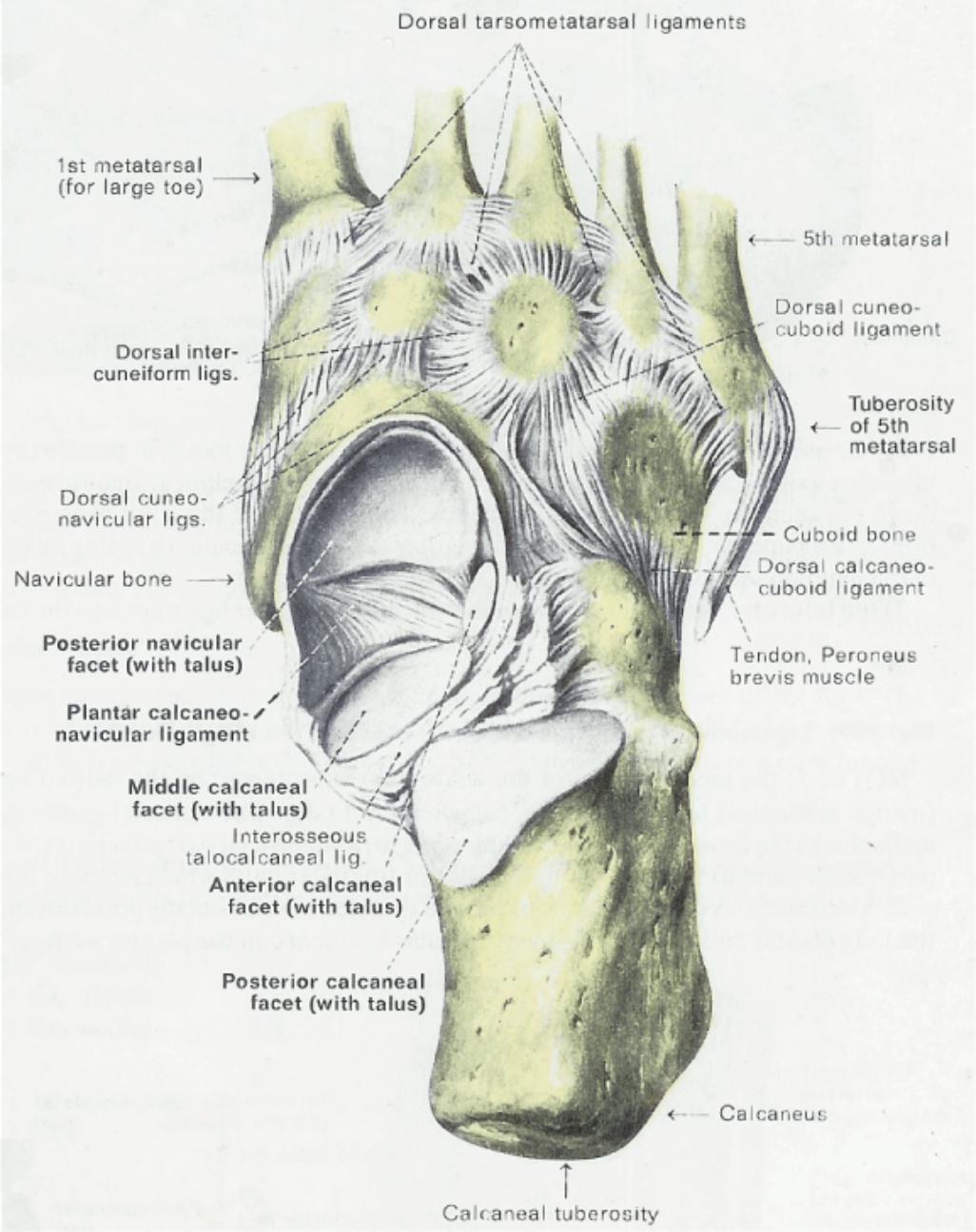
9.7 Fig 511 The Talocalcaneonavicular Joint (Viewed from above) Right
9.7.1 The talus has been removed. This reveals the three articulations it makes with the calcaneus and the anterior'articulation it makes with the navicular bone. Observe the plantar calcaneonavicular ("spring") ligament stretching across the plantar aspect of the talocalcaneonavicular joint.
9.7.2
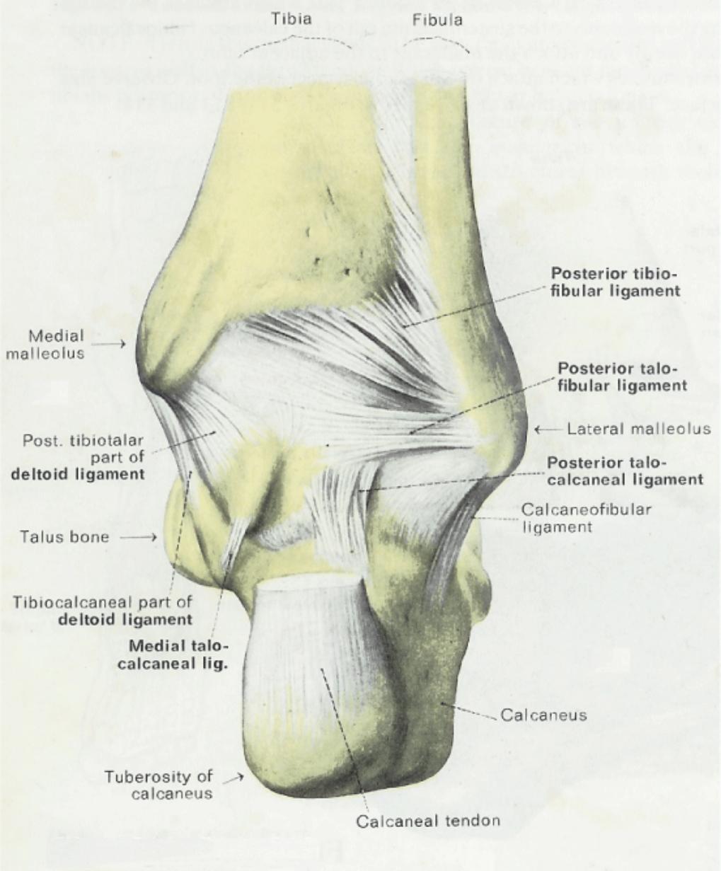
9.8 Fig 512 The Ankle Joint (Talocrural) Viewed from Behind (Right Foot)
9.8.1 The ankle joint is a ginglymus or hinge joint. The bony structures participating in this joint superiorly are the distal end of the tibia and its medial malleolus, and the distal fibula and its lateral malleolus. Together these structures form a concave receptacle for the convex proximal surface of the talus.
9.8.2 The posterior aspect of the articular capsule is somewhat strengthened by the posterior talofibular and posterior tibiofibular ligaments. Laterally, the calcaneofibular ligament and medially, the strong deltoid ligament assist in protecting this joint.
9.8.3 The ligamentous bands which help to stabilize the talocalcaneal articulation posteriorly: the posterior and medial talocalcaneal ligaments
9.8.4
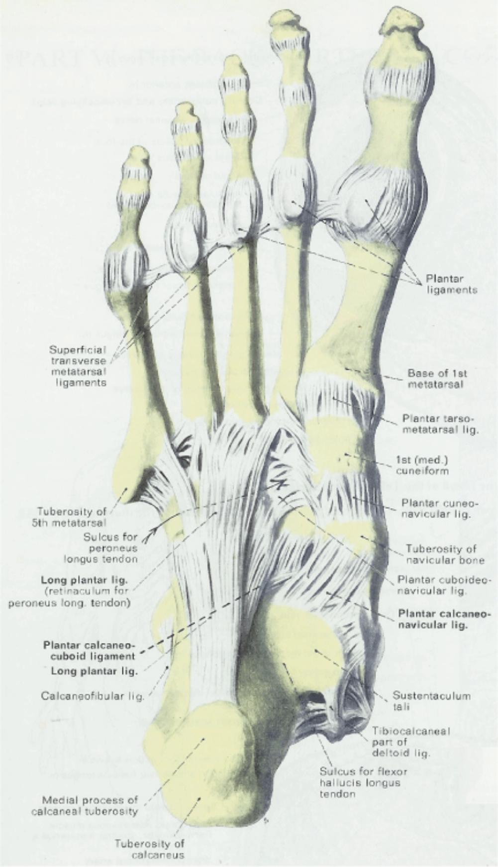
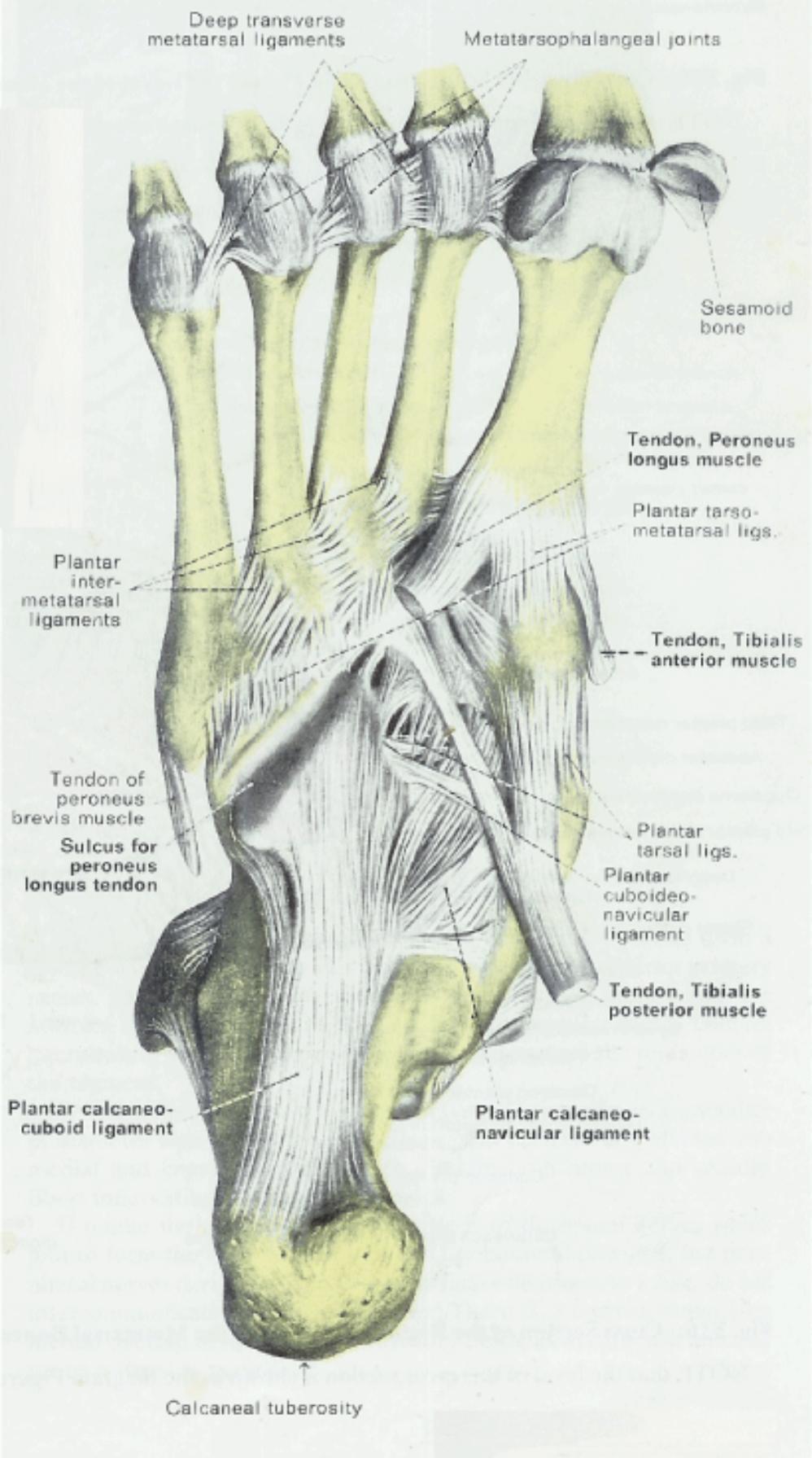
9.9 Fig 513 Ligaments on the Plantar Surface of the Right Foot (Superficial)
9.9.1 The long plantar ligament is the longest and most superficial of the plantar tarsal ligaments. It stretches from the calcaneus posteriorly to an oblique ridge on the plantar surface of the cuboid, where most of its deeper fibers terminate. A number of the more superficial fibers pass over the cuboid to insert on the bases of the lateral three metatarsal bones, thereby forming a tunnel or retinaculum for the peroneus longus tendon.
9.9.2 The plantar calcaneocuboid or short plantar ligament is very strong and lies deeper to the long plantar ligament and closer to the bones. More medially, identify the fibroelastic plantar calcaneonavicular (spring) ligament. It is attached to the sustentaculum tali of the calcaneus and extends along the entire inferior surface of the navicular bone.
9.9.3
9.10 Fig 514 The Plantar Calcaneonavicular Ligament and the Insertions of three Tendons (Right Foot)
9.10.1 The metatarsal extensions of the long plantar ligament have been cut away to reveal the groove for the tendon of the peroneus longus muscle. This tendon is seen inserting onto the base of the 1st metatarsal bone. It also sends a small slip of insertion to the 1st cuneiform. Two other long tendons inserting on the medial side of the plantar surface are those of the tibialis anterior and tibialis posterior muscles.
9.10.2 That the fibers of the calcaneocuboid (short plantar) and calcaneonavicular (spring) ligaments all stem from the calcaneus and then diverge in a radial manner toward the medial side of the foot. Observe that the course and insertion of the tibialis posterior tendon also lends some support to the short tendon and spring ligaments.
9.10.3
9.11 Synovial Joint Types
Back
Table of Contents References
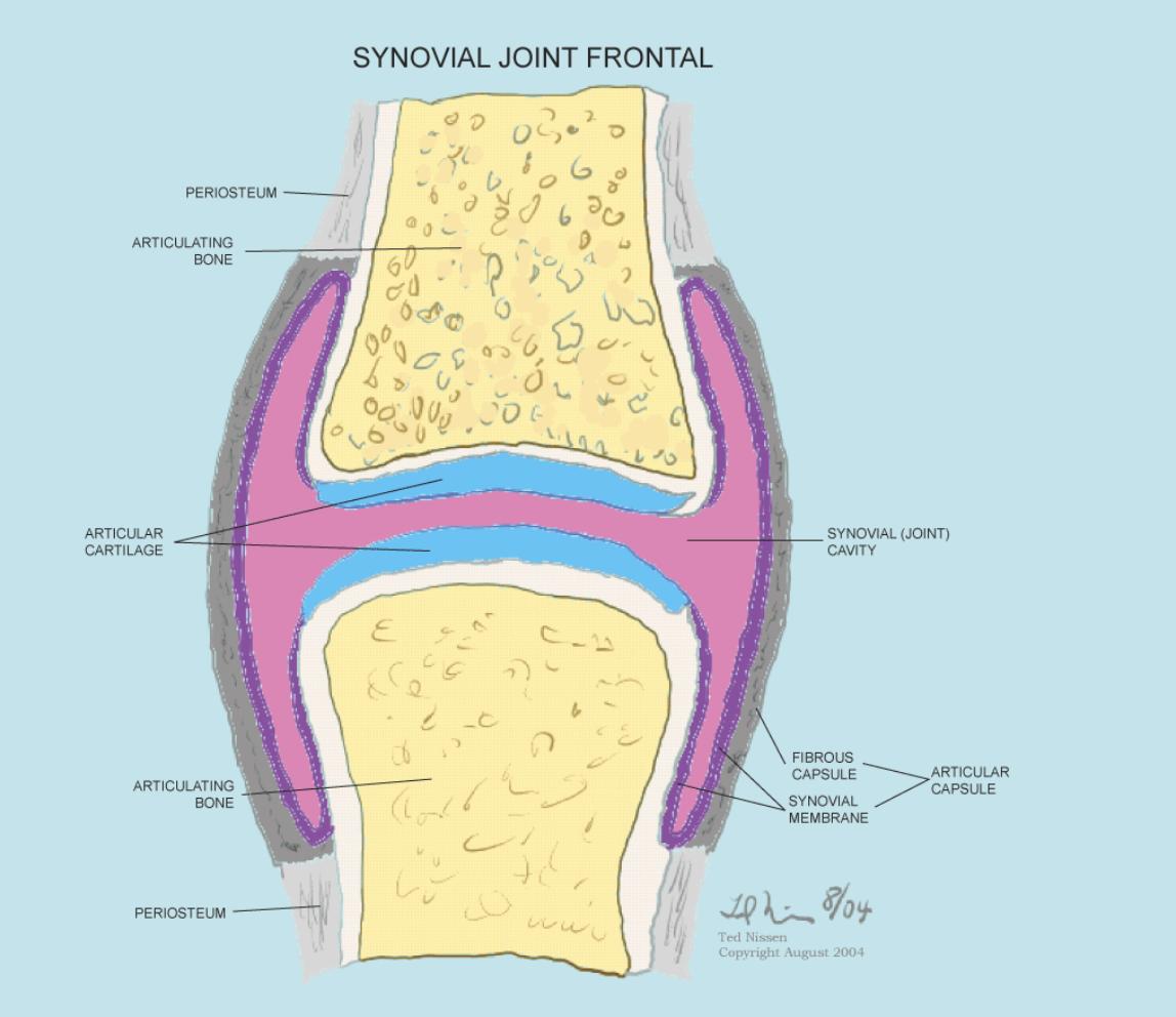
9.12 Generalized Synovial Joint Capsule Frontal Section
Back
Table of Contents References
10 Muscle Tissue
Back
Table of Contents References
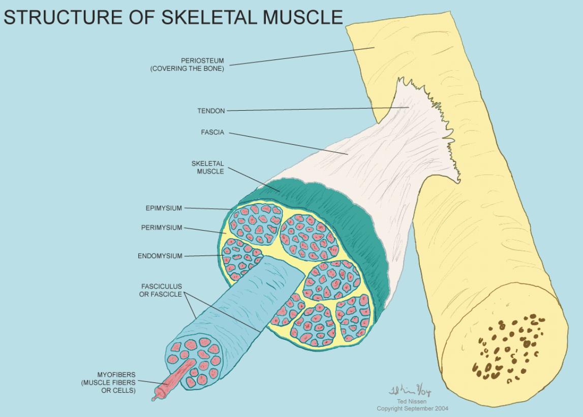
10.1 Structure of Skeletal Muscle
Back
Table of Contents References
10.2 Connective Tissue Layers
Back
Table of Contents References
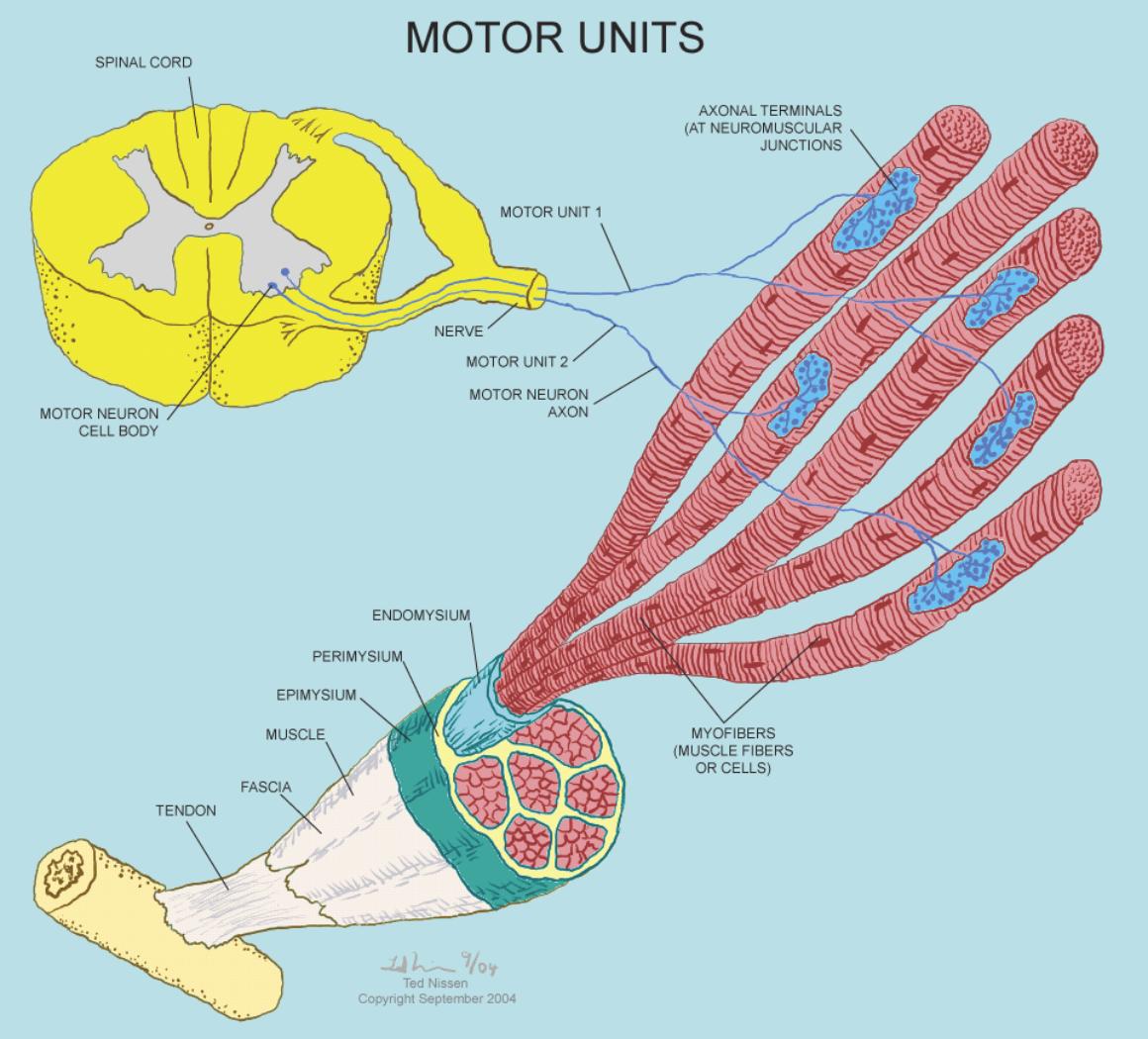
10.3 Motor Units
Back
Table of Contents References
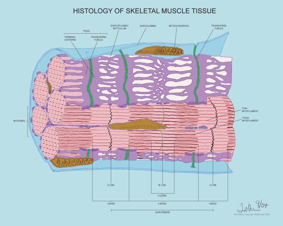
10.4 Histology of Skeletal Muscle Tissue
Back
Table of Contents References
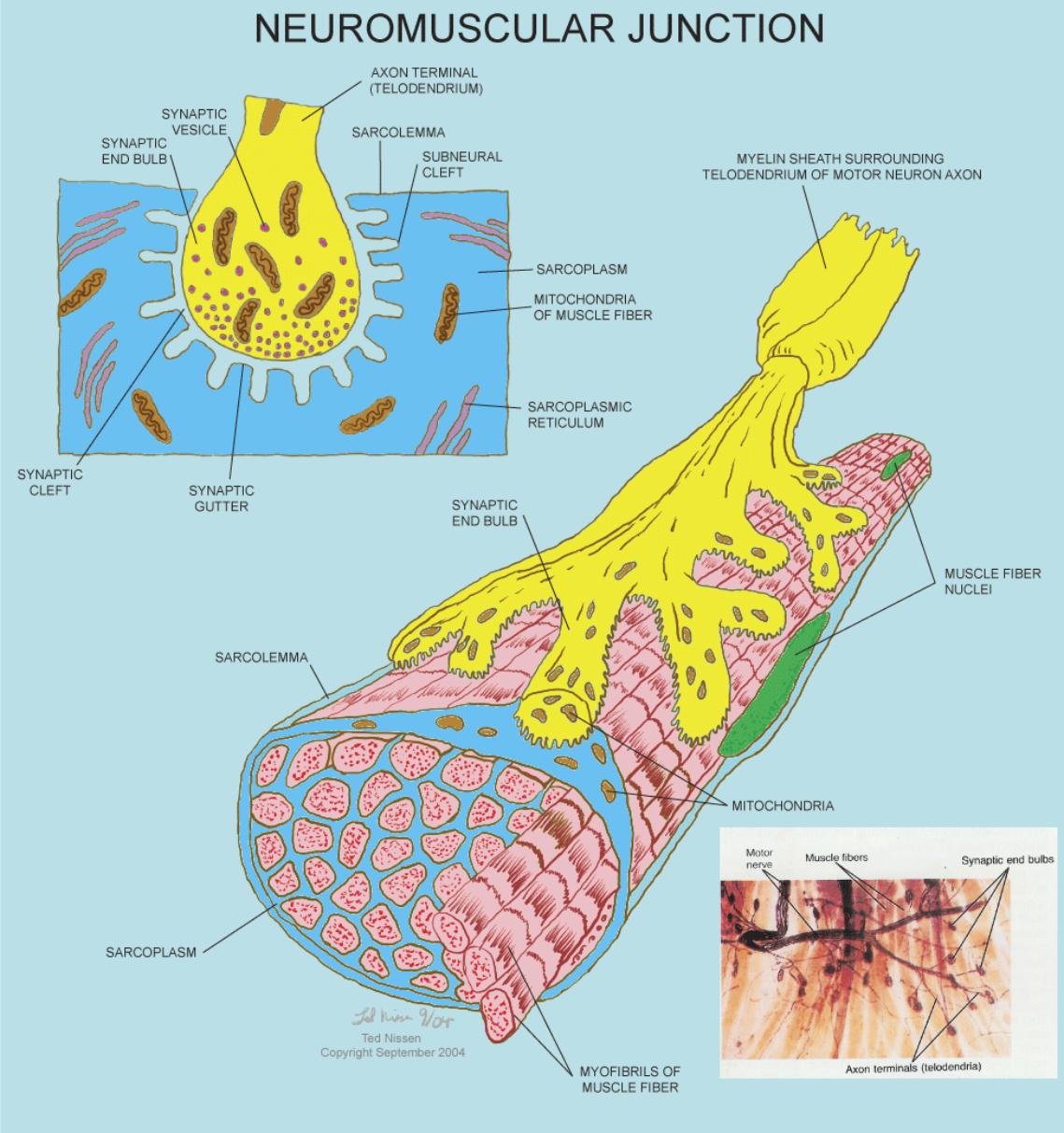
10.5 Neuromuscular Junction
Back
Table of Contents References
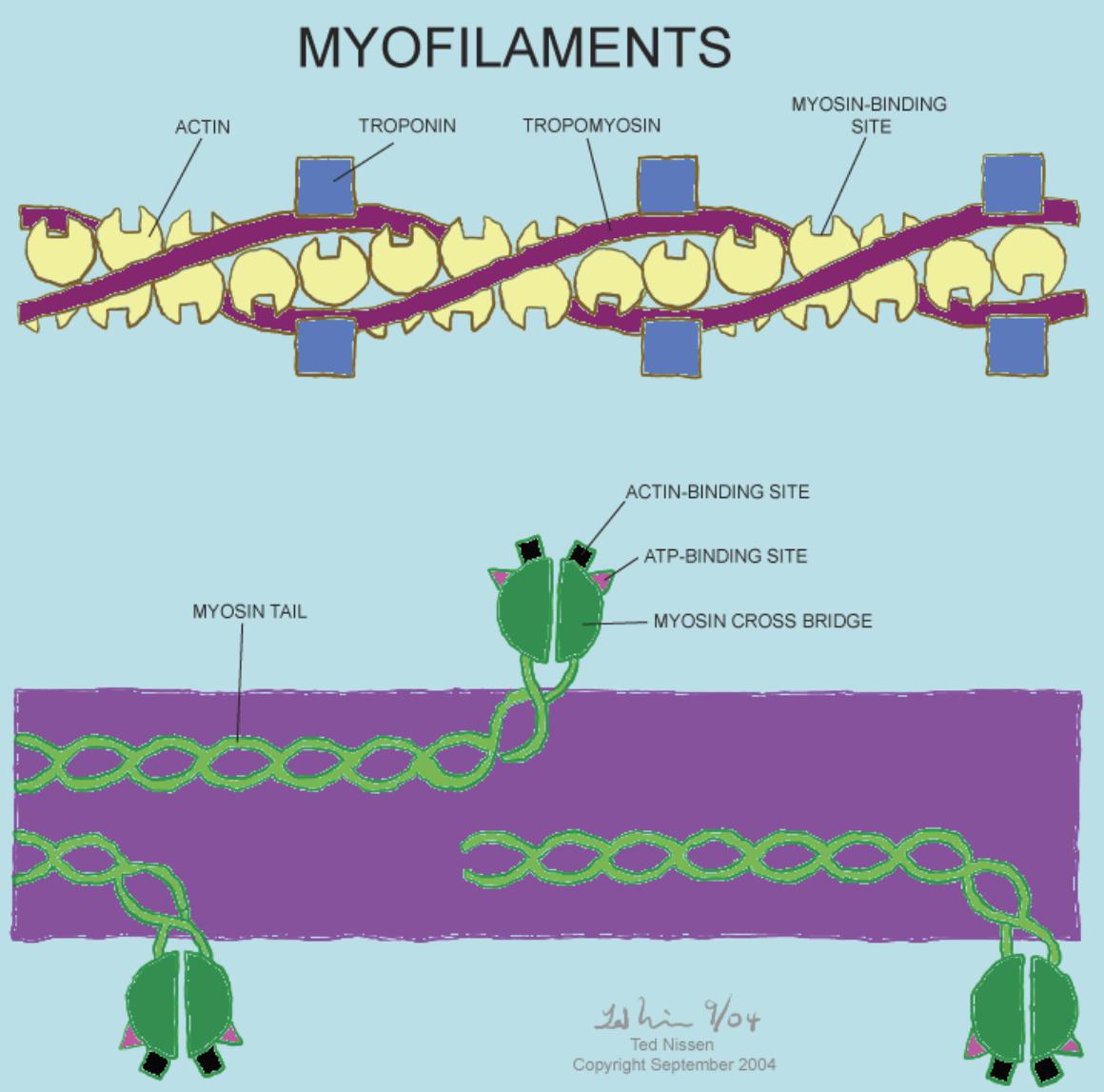
10.6 Myofilaments Detailed Structure
Back
Table of Contents References
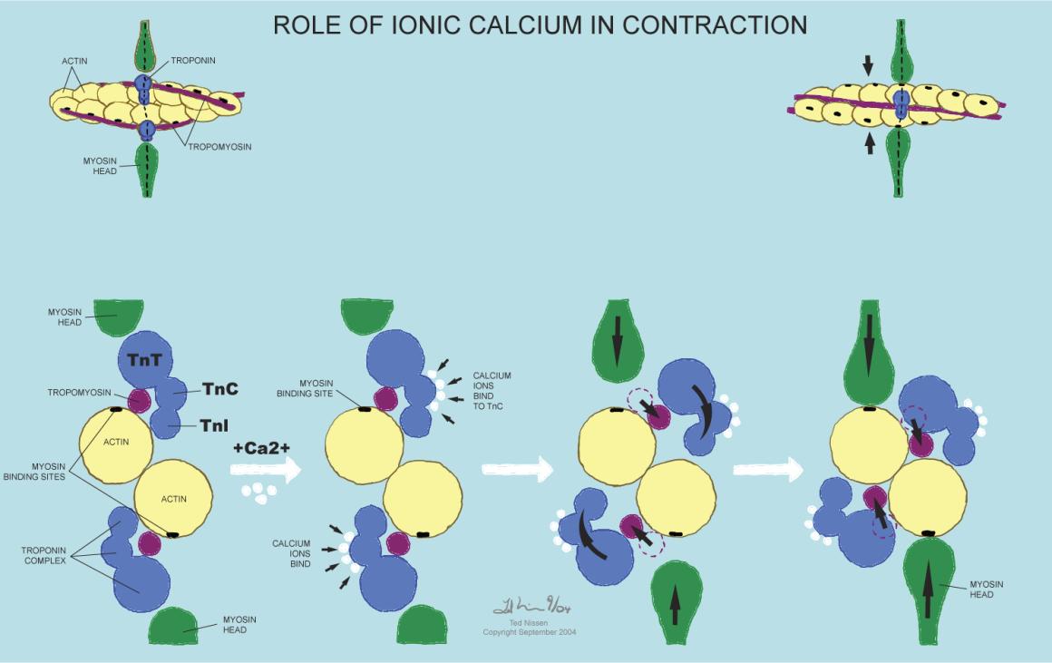
10.7 Role of Ionic Calcium in Contraction
Back
Table of Contents References
10.8 Event Sequence Actin Filament Sliding
Back
Table of Contents References
10.9 Sequence of events in Excitation-Contraction Coupling
Back
Table of Contents References
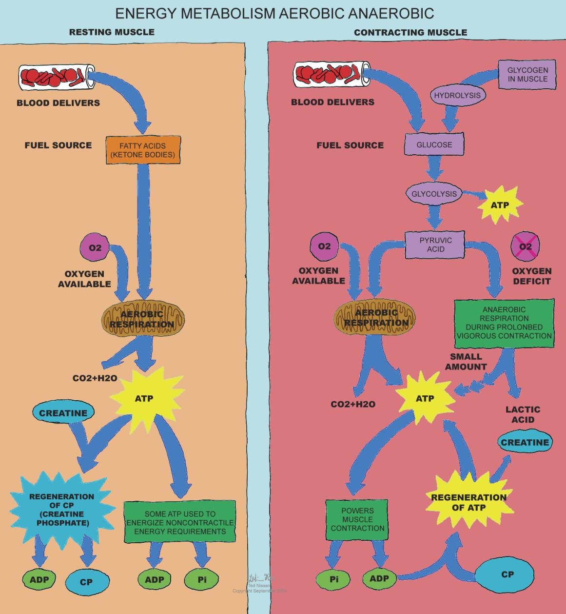
10.10
Skeletal Muscle
Energy Metabolism Aerobic Anaerobic
Back
Table of Contents References
11 Muscular System
Back
Table of Contents References
11.1 Classes of Levers
Back
Table of Contents References
12 Nervous Tissue
Back Table of Contents References
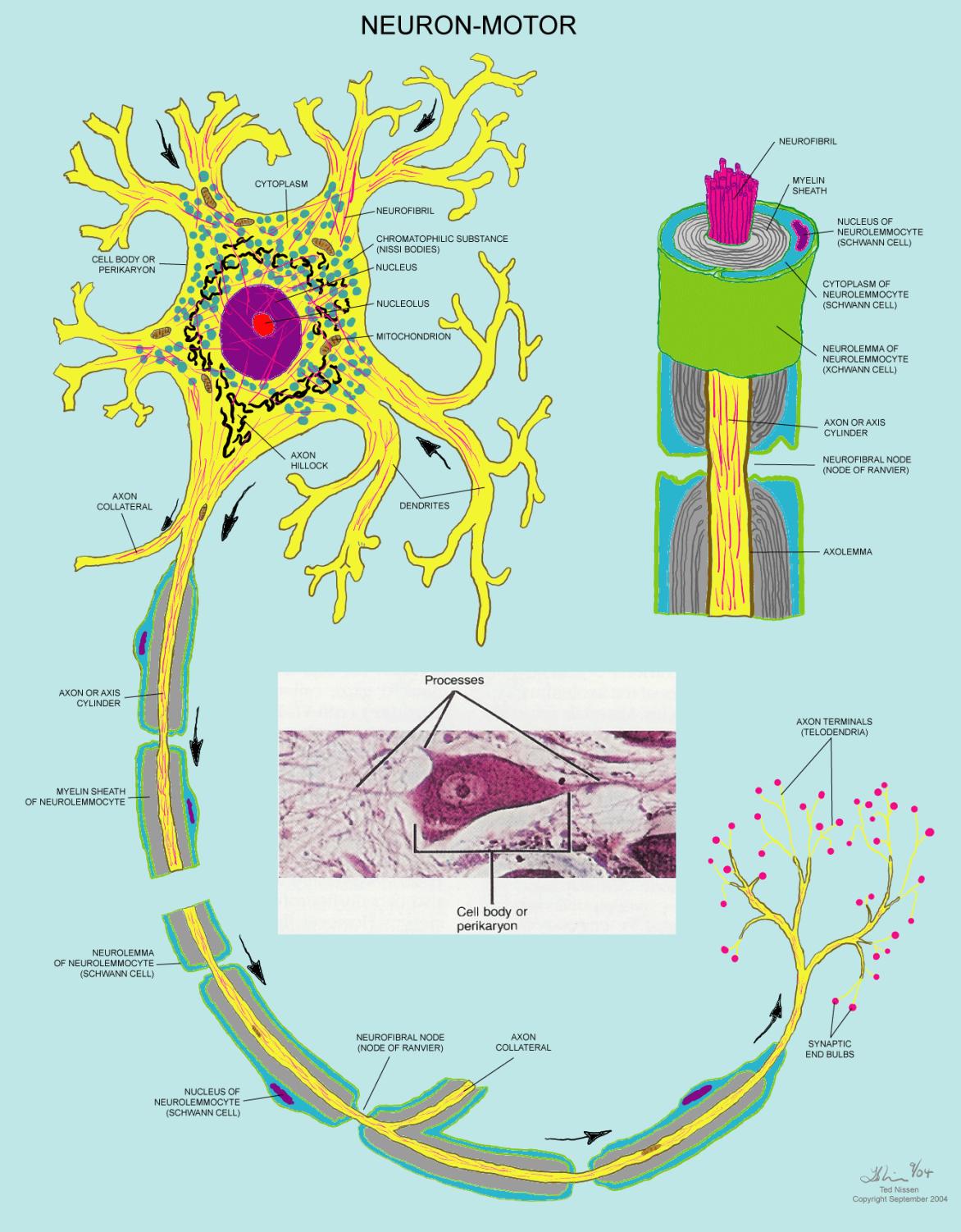
12.1 Neuron-Motor
Back
Table of Contents References
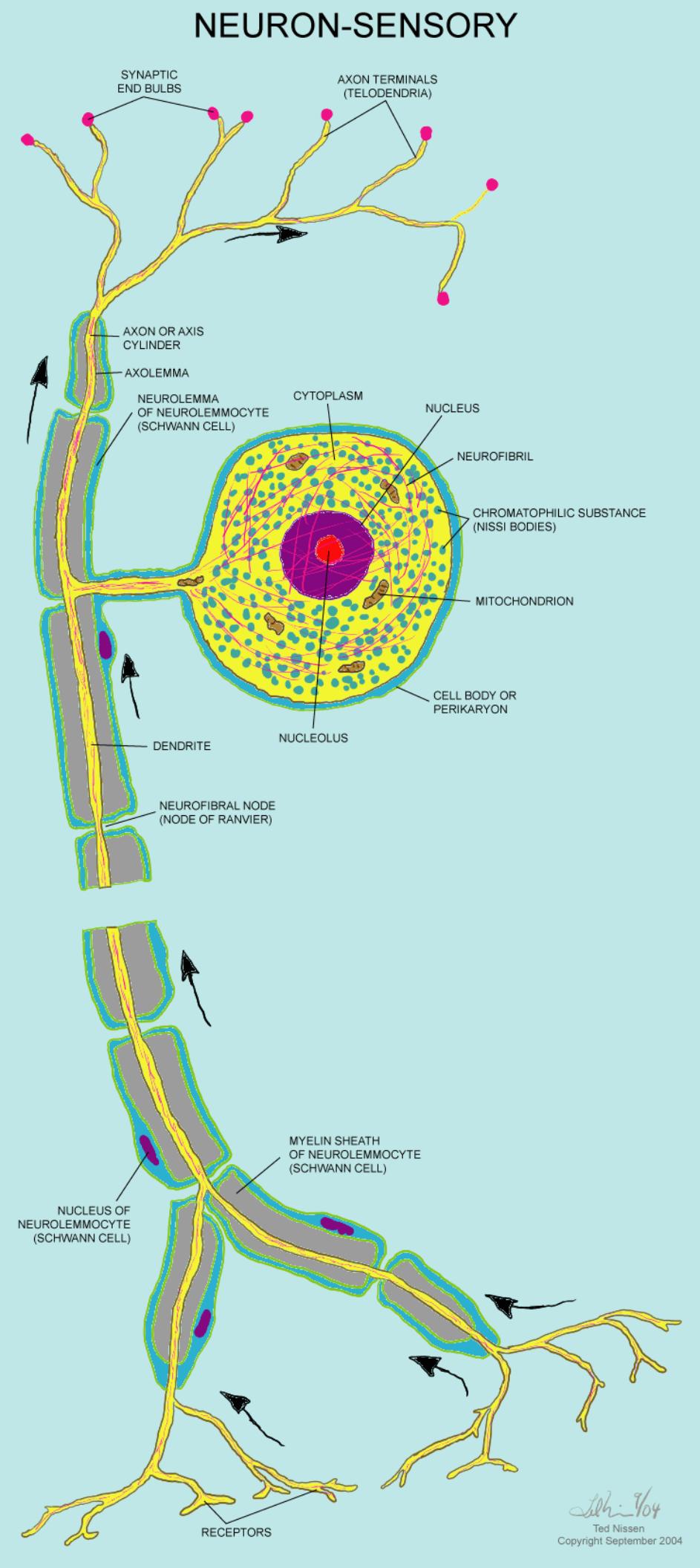
12.2 Neuron-Sensory
Back
Table of Contents References
13
Spinal Cord and Spinal Nerves
Back
Table of Contents References
13.1 Exit of Spinal Nerves
Back
Table of Contents References
13.2 Cutaneous Nerve Distribution Plantar Foot & Posterior Lower Leg
Back
Table of Contents References
13.3 Lumbar and Sacral Plexus
Back
Table of Contents References
13.4 Nerve Distribution (Lumbar and Sacral Plexus)
Back
Table of Contents References
13.5 Stretch Reflex-Muscle Spindles
Back
Table of Contents References
13.6 Tendon Reflex-Golgi Tendon Organs
Back
Table of Contents References
14 The Brain and Cranial Nerves
Back
Table of Contents References
15 Sensory, Motor, and Integrative Systems
Back
Table of Contents References
16 Autonomic Nervous System
Back
Table of Contents References
-
17 Special Senses
Back
Table of Contents References
18 Endocrine System
Back
Table of Contents References
19 Cardiovascular System-Blood
Back
Table of Contents References
20 Cardiovascular System-Heart
Back
Table of Contents References
21 Cardiovascular System-Vessels and Routes
Back
Table of Contents References
21.1 ARTERIES OF PELVIS & RIGHT LOWER EXTREMITY ANTERIOR/POSTERIOR
Back
Table of Contents References
21.2 VEINS OF PELVIS & RIGHT LOWER EXTREMITY ANTERIOR/POSTERIOR
Back
Table of Contents References
22 Lymphatic and Immune System
Back
Table of Contents References
23 Respiratory System
Back
Table of Contents References
24 Digestive System
Back
Table of Contents References
25 Metabolism
Back
Table of Contents References
26 Urinary System
Back
Table of Contents References
27 Fluid, Electrolyte, and Acid-Base Dynamics
Back
Table of Contents References
28 Reproductive System
Back Table of Contents References
29 Development and Inheritance
Back Table of Contents References
30 KEEPING THIS SPACE WARM