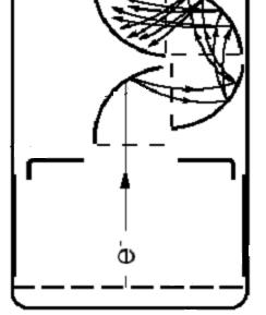
Figure 1
Ultra-weak Photon Emission from Human Hand: Influence of
Temperature and Oxygen Concentration on Emission
Kimitsugu Nakamura,1 Mitsuo Hiramatsu*2
1 Electron Tube Division, Hamamatsu Photonics K.K., Toyooka, JAPAN and
2 Central Research Laboratory, Hamamatsu Photonics K.K., Hamakita, JAPAN
*To whom correspondence should be addressed at: Central Research Laboratory, Hamamatsu Photonics K.K., Hirakuchi, Hamakita, 434-8601, JAPAN, Phone: 81-53-584-0250; Fax: 81-53-584-0260; e-mail: hiramatu@crl.hpk.co.jp
Key words: ultra-weak photon emission, photon-counting, human hand
Abbreviations: UPE, ultra-weak photon emission; PMT, photomultiplier tube;
Abstract
We have studied ultra-weak photon emission (UPE)
from living organisms. We report here
some features of the UPE from human hand by means of photon counting
techniques. The intensity of the UPE
depended on the position of human hand; nail > finger > palm. As the temperature declined, the intensity
of the UPE from the palm decreased.
Further, as oxygen concentration around the palm was lowered, the
intensity of the UPE from the palm decreased.
These results show the UPE from the palm partly
contains emissions based on oxidation reaction on skin surface as a potential. When we used mineral oil between the
photomultiplier tube and the palm, the intensity of the UPE increased twice as
much, which indicates the UPE from the inside of the skin certainly exists. The fact may be explained by refractive
index matching. As mentioned above, we considered the generation mechanism
of photons emitted from the human hand.
Many living organisms are known to emit ultra-weak photon emission (UPE), referred to as "biophotons" [1,2]. This is different from the bioluminescence with high quantum efficiencies. The existence of the UPE was not known before the 1950s because of its extremely weak intensity (i.e., 10 to 104 photons s-1 cm-2), but it has been demonstrated by means of photon counting techniques [3]. UPE is believed to be strongly related to mechanisms of life phenomena, but at the present time almost all of the phenomena associated with the UPE are poorly understood, including their mechanisms.
Based on the development of a two-dimensional imager with ultra-high sensitivity, we reported two-dimensional imaging of the UPE from intact soybean roots [4]. As an application of the use of the UPE, we were able to obtain information about the defense response of the sweet potato to infection by the non-pathogenic fungus Fusarium oxysporum [5,6].
The UPE from human hands and a forehead was reported over a period of nine months by Cohen and Popp [7]. We report here some features of the UPE from human hand, influence of temperature and oxygen concentration on the UPE. These results show the UPE from the palm partly contains emissions based on oxidation reaction not only on the skin surface but also inside the skin as a potential.
<<Fig.1>>
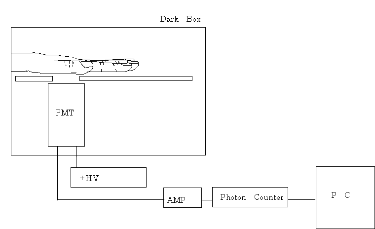
To observe the time-dependent intensity variation in the UPE, a C2550 photon counter (Hamamatsu Photonics K.K.) with a built-in, high-voltage, stabilized, direct-current power source was used. The device counts the number of photons detected by R647 (1/2 inch), R331 (2 inch), and R329 (2 inch) photomultiplier tubes (PMT, Hamamatsu Photonics K.K.) with bialkali photocathode, providing a spectral response from 185 to 650 nm. Two poles were set in the dark box to reproducibly fix the position of the palm, one was between the thumb and the forefinger, and the other was between forefinger and middle finger as shown in Fig.1. To measure the surface temperature of the palm, a sensor (Thermo Recorder RS-11, Takai Espec Corp.) was used. A commercially available PET (polyethylene terephthalate) bottle containing hot or cold water was used for the experiments of temperature influence on the UPE. After grasping the PET bottle for about 10 seconds, the UPE from the warmed or cooled hand was measured at once in the dark box. The UPE from a rubber glove (As One Corporation) in place of the human hand was measured as a control experiment with inanimate object. Nitrogen or oxygen gas from a compressed gas tank was introduced into the dark box without purification to control the concentration of oxygen. Mineral oil (Intestinal Lubricant Manufactured For shire US, Inc.) with refractive index of 1.49 was used as received.
3.1 Influence of
temperature on the UPE
It was found that the intensity of the UPE depended on the positions of the hand, nail > finger > palm by the PMT (R647) with 1/2 inch photocathode of dark 40 counts s-1 (background). About 100 counts s-1 per the photocathode area for the nail was observed as total of UPE signal 60 counts s-1 and dark 40 counts s-1 (background). Total counts for the finger and the palm were ca. 80 counts s-1 and ca. 60 counts s-1, respectively. In order to obtain stable and reproducible data, we decided the measurement position to the palm. In the case of the measurement of the palm, the PMT (R331) with 2-inch photocathode was used to collect photons with lower intensity. It is very important to use a PMT with larger area of photocathode for the UPE with very low intensity, because detecting as much signal as possible is advantageous for improving signal to noise ratio. In the case of area light source such as the human hand, especially, the PMT with larger area of photocathode is favorable to obtain higher signal to noise ratio, because it is the most important to realize larger solid-angle and to collect many photons for the area light source with the UPE.
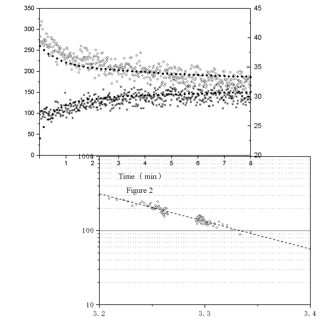
Temperature dependence of the UPE from the palm was measured using the photon counting method. When the hand was warmed at 38.6 ℃ with hot water, the intensity of the UPE increased. As the temperature gradually declined, the intensity of the UPE from palm decreased as shown in Fig.2 (a). When the hand was cooled at 22.9 ℃ with ice water, the intensity of the UPE decreased. As the temperature gradually rose, the intensity of the UPE from palm increased as shown in Fig.2 (b). From these data, we can calculate the activation energy of about 0.7 eV with coefficient of correlation of 0.94 (Fig.3). However, these data include the temperature dependence of dark counts of PMT. The dark counts of the PMT changed from ca. 35 counts s-1 to ca. 50 counts s-1 between 22.9 ℃ and 38.6 ℃. With consideration for the temperature dependence of dark counts of PMT, the activation energy of about 0.6 eV can be calculated (0.7 eV minus 0.1 eV). Namely, these findings suggest the UPE is a kind of chemiluminescence, which is luminescence based on a chemical reaction with the activation energy of about 0.6 eV. The UPE from a rubber glove containing warm or cold water in place of the human hand was measured as a control experiment with inanimate object, but the experiments was not successful because of too strong emission.
<<Fig.2>>
<<Fig.3>>
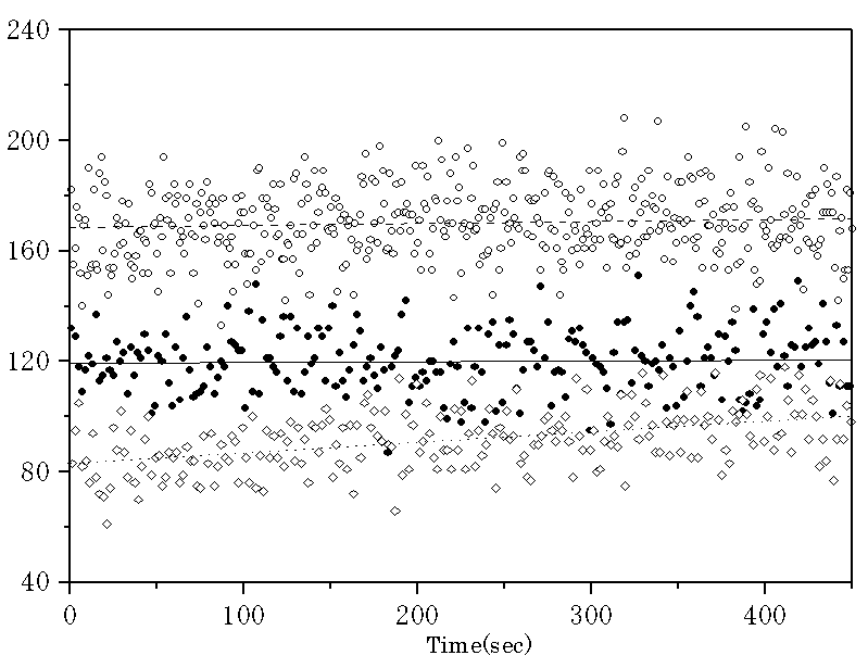
First, the intensity of
the UPE was measured under air (about 21% oxygen) at room temperature as the
control experiment using photomultiplier tube R331 (Fig.4 (a)). About 85 counts s-1 was obtained
(120 counts s-1 observed minus 35 counts s-1 background)
as shown in Fig.4 (a). Next, a lot of nitrogen
gas was introduced into the dark box to create an environment with almost no
oxygen gas. Then the intensity of the
UPE signal decreased around 50 counts s-1 (85 counts s-1
observed minus 35 counts s-1 background) as shown inFig.4 (b). This fact shows the UPE includes the
oxidation on skin surface by oxygen.
Roughly speaking, about 40% of the UPE is based on the oxygen oxidation
on the skin surface. The rest may be
from the inside of the skin. In order
to prove the hypotheses, much oxygen gas was introduced into the dark box to
fill oxygen gas around palm. As we
expected, the intensity of the UPE signal increased around 140 counts s-1
(175 counts s-1 observed minus 35 counts s-1 background)
as shown in Fig.4 (c).
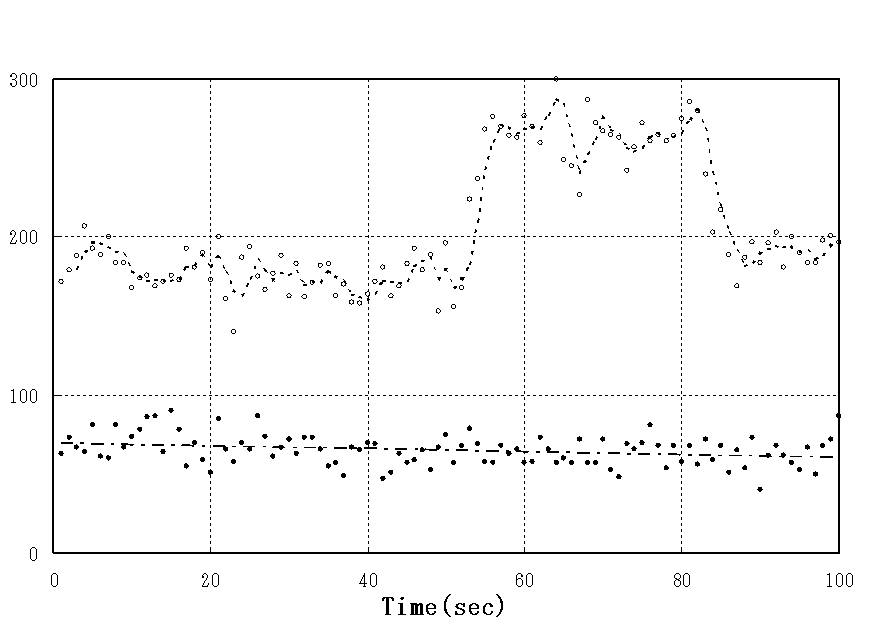
In addition, we found
out how to increase the UPE. In the
presence of an air layer, about 100 counts s-1 (180 counts s-1
observed minus 80 counts s-1 background) was obtained, while in the absence of air layer by contact of PMT (R329; 2 inch)
with palm through mineral oil, about 200 counts s-1 (280 counts s-1
observed minus 80 counts s-1 background) was obtained as
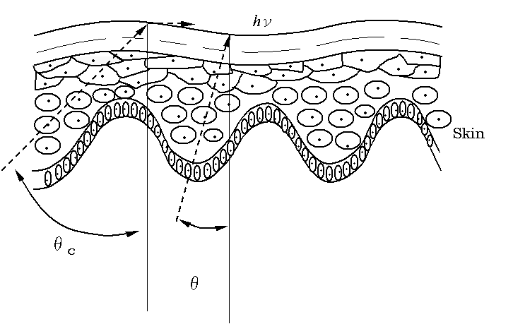
shown in Fig.5. As the refractive index
of the mineral oil is 1.49 as same as that of the skin, the matching of the
refractive index will bring about the increase twice as much as shown in
Fig.6. In order to verify our
hypothesis, water with the refractive index of 1.33 was used in place of the
mineral oil. In the absence of air
layer by contact of PMT with palm through water, about 200 counts s-1 (280
counts s-1 observed minus 80 counts s-1 background) was
0btained. In addition, the contact
without the mineral oil or water of the hand with the glass of the PMT gave us
the similar results. Namely, the
absence of air layer is the most important to obtain many photons. These results indicate that the contact of
the hand with the oil does not lead to an increase of the photon emission of
the hand by a chemical reaction and that the emission from the inside of the
skin certainly exists. But the total
intensity increased of the UPE includes a part of the dark counts increased by
the contact of the hand with the PMT.
In summary, ultra-weak
photon emission (UPE) was detectable without cooling from the living organism
using photo-multipliers manufactured by Hamamatsu Photonics. About 40% of the UPE is considered to be
oxidization by the oxygen on the surface of the skin, and it is considered that
remaining 60% is from the inside of the skin. It is expected that the UPE from this inside of the skin is based
on the oxidation-reduction reaction inside a living body. We believe that the temperature dependency of
the UPE is partly based on an oxidation-reduction reaction. An activation energy of about 0.6 eV is
obtained from this temperature dependence curve.
The UPE is spontaneous
emission without excitation from the outside.
So the UPE has some potential of applications for non- invasive
diagnosis. Additional studies are needed to elucidate the entire mechanisms for
the UPE.
<<Fig.4>>
<<Fig.5>>
<<Fig.6 >>
Acknowledgements―We thank Dr. Keith Bennett for helpful discussion and
careful correction of the manuscript.
REFERENCES
[1] D. Slawinska, J, Slawinski, Biological
chemiluminescence, Photochem.
Photobiol., 37 (1983) 709-715.
[2] H. J. Niggli, Ultraweak photons emitted by cells: biophotons, J.
Photochem. Photobiol. B: Biol., 14 (1992) 144-146.
[3] L. Colli, U. Facchini, G. Guidotti, R. Dugnani Lonati,
M. Orsenigo, O. Sommariva, Further measurements on the bioluminescence of the
seedlings, Experientia, 11 (1955)
479-481.
[4] T. Ichimura, M. Hiramatsu, N. Hirai, T. Hayakawa,
Two-dimensional imaging of ultra-weak emission from intact soybean roots, Photochem. Photobiol., 50 (1989) 283-286.
[5] T. Makino, K. Kato, H. Iyozumi, H. Honzawa, Y. Tachiiri,
M. Hiramatsu, Ultraweak luminescence generated by sweet potato and fusarium
oxysporum interactions associated with a defense response, Photochem. Photobiol., 64 (1996) 953-956.
[6] M. Hiramatsu, in J. J. Chang, J. Fisch, F.-A. Popp (Eds),
Biophotons, Kluwer Academic Publishers, Netherlands, 1998, pp. 45-55.
[7] S. Cohen, F. A. Popp, Biophoton emission of the human
body, J. Photochem. Photobiol. B: Biol., 40 (1997) 187-189.
Figure
1. Schematic drawing for measuring ultra-weak luminescence from human hand
Figure
2. Influence of temperature on the ultra-weak photon emission
(a) after the hand was warmed, ◆, skin
temperature, and ○, photon emission
(b) after the hand was cooled, ●, skin
temperature, and *, photon emission
Figure
3. Arrhenius plot for Influence of temperature on the ultra-weak photon
emission
Figure
4. Influence of oxygen concentration
on the ultra-weak photon emission
(a) ■, under air, (b) ◇, under nitrogen gas, and
(c) ○, under oxygen gas
Figure
5. Enhancement of the ultra-weak
photon emission by refractive index matching
○, photon emission from
hand; ●, dark counts of photomultiplier tube (background)
Figure 6. The course of the light from the inside of human
hand (skin)
θc, critical angle = 42°; θ, angle reaching to
photomultiplier tube, 0≦θ<θc
Figure 1


(1/T)×10-3 ( sec ) s


Photomultiplier Tube e-
![]()
![]()