
Ultra-weak Photon (Biophoton ) Emissions
(UPE)-Background Information
By
Ted Nissen M.A. M.T.
Copyright © September 2006 Ted Nissen
Articles & Abstracts Discussed
http://www.anatomyfacts.com/Muscle/photonr.html
Bibliography
http://www.anatomyfacts.com/research/photonrb.htm
Review of Literature
http://www.anatomyfacts.com/research/photonr.htm
Scientific Method
http://www.anatomyfacts.com/Muscle/scientificm.htm
Introduction
Basic Physics and Chemistry
I wish I had
paid more attention in my high school physics and chemistry classes but instead
I counted ceiling tiles, wrote bad poetry and picked at my zits. With that in
mind I will try to explain what I remember about photons, physics and chemistry
in general Chemical Organization .
What follows could have factual errors so beware. About 4.5 billion years (that
is approximately 4500 million years-hard to imagine) ago our Sun formed as a
result of hydrogen atoms (there are 118 elements of which 92 are naturally
occurring. Periodic Table These are
unique atoms which are detailed in the elemental table) compressing so much
that the relatively weak electrical force exerted by the electrons (Like
negative charges repel) of the hydrogen atoms could no longer oppose one
another. Remember an atom is composed of electrons (-charge), which move in
fixed orbits around the central nucleus, which contains protons (+ charge) and
neutrons (neutral charge). The protons are held together by the strong nuclear
force of the neutrons otherwise because like charges repel they would fly apart
disintegrating all matter. Electrons (- charge) are held in their orbits around
the protons (+ charge) because opposite charges attract. Likewise electrons
normally repel adjacent atoms so that atoms don’t normally dissolve into one
another. This is considered a relatively weak electrical force, which is a good
thing because then under the right circumstances atoms can combine to form new
elements. Chemistry studies the various atomic combinations. It’s almost like legos
the childhood play construction game. 99% of the universe is comprised of
hydrogen (H) and helium (HE). This is good because they are the simplest atoms
with one and two electrons orbiting their 1 and two protons and neutrons
respectively. Simple atoms can then be used by the compressive forces in the
sun and extreme heat to form a host of new elements. Most of the other 90
naturally occurring elements are made in stars. We are mostly made of star
stuff.
Because there is
so much hydrogen floating around in space over time (millions of years) it
becomes compressed due to the gravitational attraction of matter. Eventually
the hydrogen atoms collapse into one another (Fusion) to form helium. When that
happens photons are produced to form visible and invisible light. Photons are
thus produced as a result of chemical reaction when electrons orbits degrade or
when electrons are lost. It is the reason you see sunlight and it is still
going on today. Photons take about 8 minutes to get from the sun to earth traveling
at the speed of light at about 186,000 miles per second. Photons generally
bounce off things and so your retina is sensitive to them and you can see
objects in your environment. When the sun runs out of hydrogen then our sun
will literally burn out (probably in about 12 billion years). A photon is a sub
atomic particle (or string). According to Edward Witten Edward Witten (M Theory) (Many physicists think he is the
smartest man alive-even smarter than Einstein) a string is a vibrating string
(think violin) and or membrane of energy. The frequency (how many times it
vibrates in a given period of time) and amplitude (How forceful the vibration
is) will determine what type of sub-atomic particle it is (quark, gluon,
photon, ect). There are 21+ sub-atomic strings (particles) (things that are
smaller than an atom). Particle Physics They can
be compressed into a very small space. When massive suns die all of their
particles collapse to form a “BLACK HOLE.” It is thought that the entire
universe that exists today is a result of these particles being compressed into
a space smaller than the size of the nucleus of one atom. This concentrated
matter then exploded into what is popularly described of as the “BIG BANG” to
form the visible universe about 14 billion years ago. It used to be thought
(Democritus (450BCE-?)) that the atom was the smallest unit of matter. Then we
began smashing atoms into one another at high speeds at which point we could
see some of these smaller particles or strings. For example, when you smash up
protons and neutrons you get quarks. Other particles such as photons can be
produced thru chemical reactions, which produce new chemical elements.
Photons
A photon could
be visualized as a tortilla or pizza pie without the topping. Throw it in your
imaginary air space and slow mo its free fall so that you can carefully observe
its properties. Notice that it is not perfectly flat because when you threw it
in the air it slid off of your hand and began undulating. That is visualize
your tortilla with waves coursing across its surface in its free fall. These
are just like waves in the ocean, which you could watch splashing onto shore
with an almost rhythmic chant. The regularity of the waves over a given period
of time could be counted. This is known as the frequency of the waveform. How
big the wave is known as the amplitude of the wave. All photons have the same
frequency and amplitude of their waveform. Instead of a tortilla you could
substitute a rubber band like string that surrounds a membrane. You could also
imagine (for our metaphorical purposes only) that the string and membrane are
made of energy. Other subatomic strings (particles) as aforementioned vibrate
at different frequencies and amplitudes but all subatomic strings are made of
the same energy. Think about that. Although string theory, is interesting it is
far from certain because we just don’t have the equipment to actually see a
vibrating string or membrane. These are elegantly elaborated mathematical
models which suggest but do not prove an almost Alice in wonderland world.
Photons
themselves also travel along electromagnetic waves. [1]
This means that visible light for example is both a particle (string) and a
wave. This was a huge debate in physics for the longest time. Sir Isaac Newton
(1643-1727) [2] believed
that light consisted of a stream of particles, while Newton’s colleagues, most
notably the Dutch physicist Christiaan Huygens (1629-1695)[3],
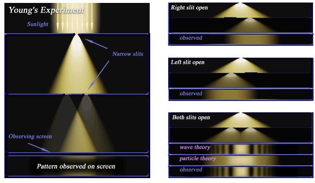
disagreed with him and argued that light is a wave. In an experiment by Thomas
Young (1773-1829) [4]
performed around 1805 known as the Double-slit experiment or two-slit
experiment [5]
the debate was settled. It’s a simple experiment that does not require an
understanding of quantum mechanics but once its implications are carefully
considered disproves Newton’s notion that light is composed of particles. Take
a single light source, cut two slits in a board, place a screen in back of the
board so that the board is between the light and the screen. The single light
source now projects through the two slits and creates two light sources, which
project onto the screen behind. If light were a particle the light projected
onto the screen would diffuse evenly onto the background screen. If light were
a wave its properties would be similar to waves in an ocean. Imagine you are
next to a beautiful lake, which is perfectly calm, with not a ripple on its
placid surface. In fact it is so smooth you can see the high snow capped
mountains, towering above the lake, reflected onto its surface. Now with both
hands hold two stones at arm length apart and drop them into the lake
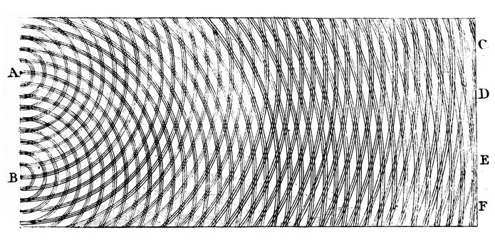
simultaneously. You will note an interference pattern where the waves of one
stone cancel out the waves of the other stone. Double-Slit Experiment Diffraction.
The areas of darkness on the screen behind the light source are the result of
the light waves interfering (Diffraction) with one another. The dark areas are
caused when peaks and troughs occur together (destructive interference) and the
light areas are caused when two peaks coincide (constructive interference).
According to this experiment, nearly 100 years after his death Newton was
proved wrong and his colleague Huygens was right. During their lives Newton and
Huygens did not know the outcome of this debate but later on both would be
proved right.
In the 20th
century Albert Einstein (1879–1955) [6],
Louis de Broglie (1892–1987) [7]
and many others postulated and confirmed that light (photons) and matter consist
of both particles and waves. This was known as the Wave–particle duality [8].
It has been shown experimentally that all-electromagnetic waves and also other
subatomic particles (strings) as well as atoms demonstrate the same
interference patterns. Photons travel at the speed of light along the
electromagnetic wave. The speed of light is 186,171.116418 miles per second
(299,792,458 metres per second (approximately 3 × 10⁸ metres per second. 1
Kilometre is 1,000 metres. 1 Kilometre is 0.621 of a mile). That means a photon
of light travels 7.48 times around the earth in one second. (Earth
circumference 40,076 km in circumference or 24,887.196 miles) The distance from
the earth to moon is 384,400 km or 238,712.4 miles so it takes light
approximately 1.28 seconds to reach the moon. Click on this link and then on
the dark image at the top of the page to see how fast light goes from the earth
to the moon in real time. Speed of Light. It takes
about 8 minutes for a photon of light to reach the earth from the sun.
String theory
was developed (Yoichiro Nambu (and later Lenny Susskind and Holger Nielsen) in
the late 1960’s and early 1970’s to explain the behavior of subatomic particles
(proton and neutron which experience the strong nuclear force). Later M-Theory
was developed in 1995 by Edward Witten to tie together the various string
theories. According to these theories photons are not really particles
(zero-dimensional point in space) but rather vibrating strings (one-dimensional
extended objects) (String Theory) [9]
and or membranes (M-Theory) [10].
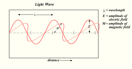
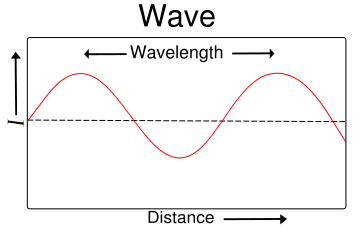
As discussed above photons move at the speed of light along a wave with a
particular frequency, wavelength and amplitude. This wave of photons is
electromagnetic radiation [11]
(light wave example)
of the electromagnetic spectrum in order of increasing frequency (radio waves, microwaves, infrared radiation,
visible light, ultraviolet radiation, X-rays and gamma rays). The frequency [12]
(Frequency Example)
is determined by counting the frequency of the wave in a given time period. The
wavelength [13] (Wave Length)
is measured as the distance between repeating units of wave pattern.
Electromagnetic radiation is actually composed of two self-propagating waves,
(one electrical-one magnetic), at right angles to each other (light wave example).
Therefore a time-varying electric field generates a magnetic field and vice
versa. Thus, as an oscillating electric field generates an oscillating magnetic
field, the magnetic field in turn generates an oscillating electric field, and
so on. These oscillating fields together form an electromagnetic wave composed
of photons traveling at the speed of light generating the electromagnetic
spectrum from radio waves to gamma rays.
Alexander
Gavrilovich Gurwitsch, also Gurvich (Russian:
Александр
Гаврилович
Гурвич 1874-1954) [14]
famous Russian embryologist, developmental biologist, medical scientist, and
Professor of Histology in Taurida University (1918-1924) discovered ultraweak
UV (260 nm) photon emissions from living tissue in the 1920’s. Prof. Gurwitsch
named these photon emissions "mitogenetic rays" (refers to UV
electromagnetic waves of photons which stimulate increased cell division
(mitosis)) because his experiments showed that they stimulated cell division
rates [15]
of nearby cells. Prof. Gurwitsch was thinking about how living tissues transfer
the information about the size and shape of organs given that chemical
reactions "do not contain spatial or temporal patterns a priori (formed or
conceived beforehand). Prof. Gurwitsch began looking for a morphogenetic (relating
to or concerned with the development of normal organic form) field, which might
regulate cell growth and differentiation. (Don’t geneticists explain this
better through DNA expression) [16]
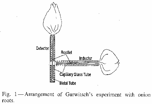
He devised what he called the basic experiment ("Grundversuch") [17].
It should be noted that normal window glass blocks UV rays and quartz glass
plate is transparent for UV light of about 260 nm. Two onion roots were
arranged at right angles to one another with the horizontal root (Inductor)
pointed towards the vertical stem (Detector) with a space for either normal
window glass or quartz glass plate (Experiment). The subject of
observation was the cell division (number of mitoses) rate on the stem where
the root tip was pointed. When window glass was placed in the space between the
root and the stem no cell division changes were noted whereas when the quartz
glass plate was placed in the space cell division (number of mitoses) increased
significantly. Prof. Gurwitsch concluded that ultraweak UV (260 nm) photon
emissions in the in the horizontal root (Inductor) were stimulating increased
cell division in the vertical stem (Detector). The lack of cell growth when a
normal window glass blocked UV stimulation and increased cell growth when quartz
glass plate facilitated UV stimulation suggested to the professor that photons
might regulate cell growth and differentiation. Prof. Gurwitsch’s work,
however, was criticized because of inaccurate photon counting methods and the
fact that cell growth can be stimulated by other forms of Electromagnetic
Radiation (radio waves, microwaves, infrared radiation, visible light,
ultraviolet radiation, X-rays and gamma rays) [18].
In addition biochemists were explaining cell growth in terms of hormones and
other biochemicals. The work of Alexander G. Gurwitsch was largely forgotten.
It is unclear whether other scientists repeated his experiments. Current
"debate surrounds such evidence and conclusions, and the difficulty of
teasing out the effects of any supposed Biophotons amid the other numerous
chemical interactions between cells makes it difficult to devise a testable
hypothesis" [19]
After World War
II in the 1940s Colli (Italy), Quickenden (Australia), Inaba (Japan) and
Boveris (USA) began experimenting with a newly devised Photomultiplier which
accurately counted single photon emissions. They all dropped Professor
Gurwitsch’s term "mitogenetic radiation" preferring the terms
"dark luminescence", "low level luminescence", "ultraweak
bioluminescence", or "ultraweak chemiluminescence". The
aforementioned researchers also proposed that these biological photon emissions
were the result of “rare oxidation (removal of electrons and hydrogen ions or
addition of oxygen) processes and radical (radicals (often referred to as free
radicals) are atomic or molecular species (a particular kind of atomic nucleus,
atom, molecule, or ion) with unpaired electrons on an otherwise open shell
configuration) reactions”.[20]
According to Popp [21]
with the exception of Quickenden (Australia), Inaba (Japan) and Boveris (USA)
the phenomenon of "low-level luminescence" “did not ever become a
serious subject of fashionable science” and was largely disregarded and
disrespected. Essentially the research by the aforementioned and other post
World War researchers regarded these photon emissions as random missteps of
cellular metabolism or as "imperfections in metabolic activity"
(Russian Biophysicist Zhuravlev & American Chemist Seliger) while
acknowledging their existence disregarded their importance.
In the 1970s
then assistant professor Fritz-Albert (Alexander-Alex) Popp (1938-Present) [22],
German Biophysicist (Earned PhD in Theoretical Physics-Mainz university) who
could be considered the modern founder of a whole new branch of biophysics
exploring Biophoton emissions, discovered a much wider spectrum of photon
emissions than had previously been recorded (200 to 800 nm). Prof. Popp coined
the term “Biophoton” and holds patents, which include the use of Biophotonics to
examine the quality of food, of the environment and in medicine, among many
others. Prof. Popp has proposed that this electromagnetic radiation
(Biophotons) is both semi-periodic and coherent but has yet to win general
approval from his colleagues.
Also in the
1970’s biochemists considered the measurement of Biophotons as a way to study
reactive oxygen species (superoxide for example) within a single cell more
specifically within the mitochondria but because biophoton production is
relatively rare within a single cell structure, overall Biophoton production
ultra-weak, and the mechanisms of production complex most biochemists were put
off. Britton Chance (1913 –Present) Eldridge Reeves Johnson University
Professor Emeritus of Biophysics at the University of Pennsylvania did measure
photon production in isolated mitochondria. But detailed subsequent studies
failed to detect a signal in dog's brain.
Hamamatsu
Photonics K.K. (founded in 1953) is a Japanese manufacturer of optical sensors,
electric light sources, and other optical devices and their applied
instruments. In the 1980’s its Electron Tube Division first developed the
Photomultiplier tube which was able to more easily and accurately measure
Biophotons. The Japanese Government began a five-year, multibillion-yen
research programme into Biophotons in 1986. Humio Inaba, an engineer at the
Research Institute of Electrical Communication at Tohoku University headed the
project.
Weak Biophoton
emissions have been discovered in everything from plant seeds to fruit flies.
Humio Inaba has noticed in study after study that distressed and diseased cells
emit significantly more photons than adjacent non-injured “healthy “cells.
These experiments have been replicated demonstrating that cell injury increases
Biophoton production. If you tear a tree leaf, for example, while measuring
Biophoton emission, a spiked rise in emission in the tens of thousands (as
opposed to a normal range of 1-1000) with what amounts to a light burst occurs.
These experiments and others have been conducted by Ken Muldrew, a biophysicist
at the University of Calgary in Alberta, Canada. In animal tissue the same
phenomena of injured cells increased photon production has also been observed.
At the Institute of Physics at the University of Catania in Italy, tumor cells
were studied. It was discovered that “mammalian tumor cells ejected photons at
rates as high as 1400 per square centimeter per minute-healthy tissues average
rates of less than 40.” [23]
Other teams of researchers have found biophoton emission from tumor cells is 4
times higher than surrounding healthy tissue.
Imaging devices
to detect disease, although still in development, are within the realm of
scientific imagination as useful non-invasive imaging tools. Reiner Vogel, a
biophysicist at the University of Freiburg in Germany, says "The emission
may give a very sensitive indication of the conditions within a cell and on the
functioning of the cellular defense mechanism," Philip Coleridge Smith, a
surgeon at University College Medical School in London, agrees. “You could
perhaps use biophotons to assess inflammation in tissues, he suggests, which
might warn of leg ulcers, for example.”
That injured
cells emit more biophotons is well established but some researchers have
suggested that biophotons may actually represent some form of communication
between cells. In the 1990’s, Guenter Albrecht-Buehler, a biophysicist at
Northwestern University Medical School in Chicago conducted experiments with
near infrared (850 nm=.850 µm-Near infrared=(0.75–1.4 µm=micrometer)) directing
light onto cell-sized latex beads, which were situated near mouse fibroblast
cells (connective tissue cells). The latex beads would project this infrared
light towards the mouse fibroblast cells. The mouse cells reached toward the
light emitted from the cell sized beads with their Pseudopodia ((false feet)
are temporary projections of eukaryotic cells). The mouse cells even began
moving towards the light source (latex beads) with some rotating 180º swiveling
and moving toward the infrared light. The power and wavelength of the light
source produced virtually no heat to direct the cellular movement or behavior.
The light alone seems the cause of the cellular behavior. If two light sources
were presented at equal intensities the cell would respond to both as if to see
two distinct light emissions. In yet another experiment Albrecht-Buehler
studied elongated hamster cells [24].
First he spread the cells onto one side of a glass pane and they grew parallel
to one another. Then he spread the cells in two thin layers on opposite sides
of a glass pane with a section in between which could accommodate a filter.
Without a filter the hamster cells grew at 45º to one another. When an infrared
filter (blocks infrared light emission from one side to the other) was added
the cells on either side of the glass pane demonstrated random orientations.
The
aforementioned and other cumulative research prompts Albrecht-Buehler to
speculate on the meaning. Perhaps this infrared light is emitted represents
cell-to-cell communication to help determine orientation, either parallel if
next to each other or criss-cross if on opposing sides. The criss cross pattern
is adaptive because it provides extra strength. Is there some kind of eye
within the cell that detects light? Albrecht-Buehler speculates that the
centrioles within the cell are potentially light sensitive because he says
their microtubule cylindrical structure creates slanted blades, which act like
blinds, allowing light in but only from certain angles. This arrangement could
act as a photoreceptor to determine which direction the photons emanate. The
microtubules-hollow filaments could act as fiber optics to direct light from
the periphery of the centrioles to the core. Are cells talking to each other?
Albrecht-Buehler guesses that embryos might signal their position with photons
and receives information for other cells to know how and where they fit into
the developing body. If this signally system like a language could be learned
could be redirect cancer cells to stop growing or enhance would healing, or
send signals to perform unforeseen tasks.
In the 1980’s,
Popp, then lecturer at the University of Marburg Germany, concluded that
cell-to-cell communication was evident in synchronous biophoton emissions
between cells without a light barrier vs. asynchronous biophoton emissions
between cells separated by an opaque barrier.
Cyril Frank
surgery professor at the University of Calgary’s medical school agrees with
Popp speculating that biophotons could trigger events in the receiver cell such
as: mitosis rate, protein expression but further research is needed before
certainty can be claimed.
Ken Muldrew, a
biophysicist at the University of Calgary in Alberta, Canada, is not convinced
that complex messages can be conveyed by biophotons arguing that increased
oxidation reactions may be conveyed but that’s all.
The practical
uses of the detection of biophoton emissions would include as aforementioned
early detection of diseases like cancer. Problem is how to ferret out random
photon emissions coming from the occasional but possibly significantly frequent
given the 1 million per second per cell reactions (15 trillion cells) and the
increased biophoton emissions produced by disease. This might affect the
ability to replicate the results of the aforementioned researchers. Barbara Chwirot, head of the Laboratory of
Molecular Biology of Cancer at Nicolas Copernicus University in Torun, Poland
states that this is a of
“reproducibility of results, even for relatively simple systems like
cell cultures," Biophotons may also be affected by enzyme activity as well
as a host of other factors as yet determined. Bottom line direct diagnosis of
disease is not a done deal and may require further technical or medical innovation.
Popp now heads
both the International Institute of Biophysics in Neuss, Germany (Scientists
interested in biophoton research) and runs Biophotonen. Biophotonen evaluates
food products to assure for example that beer does not contain harmful microbes
(Bitburger=German brewer). Chinese groups are perfecting food related biophoton
evaluation for the presence of unwanted bacteria.
Look for future
innovation in the form of cutting edge detectors,; avalanche photodiodes ect.
Classical
physics can’t explain how brains think says Scott Hagan, a
theoretical physicist at the British Columbia Institute of Technology in
Burnaby. Pierre St. Hilaire Interval Research Corp., Palo Alto, USA
Dick J. Bierman
University of Amsterdam, The Netherlands StarLab, and Brussels, Belgium would
all agree, “Consciousness implicates quantum coherent states in the brain” [25].
The question of how brain cells can function with massive communal simultaneous
coordinated synchronicity may be answered according to Scott Hagan by thinking
of biophotons as speed of light optic communicators. Quantum coherent states
are states where the wave functions of individual atoms combine to form a
coherent pattern [26].
According to Sir Roger Penrose, OM, FRS (1931-Present) is an English
mathematical physicist and Emeritus Rouse Ball Professor of Mathematics at the
University of Oxford and Emeritus Fellow of Wadham College, Orch OR
(“Orchestrated Objective Reduction”) [27],
may provide a conceptual framework to better understand brain function. Prof.
Ball thinks that these quantum coherent states are propagated by protein
structures within the cells as part of its cytoskeleton but more to the point
of this discussion they are found with the neural cell structure including the
axons the essential wiring of the brain. These thin tubes may be likened to
fiber optics and are thought to move energy about the cell, building junctions
between neurons and perhaps aide in memory retention. Hagan and Stuart Hameroff, associate director
of the Center for Consciousness Studies at the University of Arizona, are
proponents of this highly speculative theory that quantum coherence is mediated
by these intercellular structures and may in fact give rise to consciousness [28].
Experimental evidence of this according to Hagan is the effect that anesthetics
have in binding to the microtubules
"Because anesthetics make consciousness evaporate, their site of
action is important in determining the mechanisms responsible for consciousness."
Biophotons may use these microtubules as conduits for consciousness. This is
just a theory with scant evidentiary proof.
Kenneth J.
Dillon, B.A. in history from Georgetown University and a PhD in history from
Cornell University makes the fantastic claim that red blood cells have some
kind of biophotonic signaling [29].
Dillon claims that the circulatory system is involved in the reception,
transmission, and processing of electromagnetic data and acts as a “Animal
Magnetoreceptor” which can sense magnetic fields. It is hard to say how
carefully these claims can be supported by the data.
Biophotons
Biophotons
(Greek Bio=Life, Photon=Light) [30]
are photons emitted from living organisms including plants and animals.
Biophotons are not the same as Bioluminescence that are produced by many marine
(80% of marine creatures emit light) [31](Anglerfish
& Flashlight fish) and non-marine creatures (Glow Worms & Fire flies)
and is a result of a chemical reaction within the organism, which produces
photons, which are visible to the naked eye. The process of bioluminescence is
well understood by the biological sciences. Bioluminescence is due to a
"chemical reaction between ATP-the cell's energy store-oxygen and a
molecule called luciferin. Luciferin converts the chemical energy locked up in
ATP into photons of light." [32]
Biophoton emissions are very low intensity photon emissions from living
organisms, which are poorly understood, ill defined by the experts in the
field, and its “study is controversial and is not generally accepted as a
legitimate area of study by mainstream scientists” [33].
Does this mean that research on Biophotons is accepted in mainstream journals?
This is a specialized area of biophysics known as Biophotonics (Popp), which
involves the study of the relationship between biological materials and the
emission of photons. It “refers to emission, detection, absorption, reflection,
modification, and creation of radiation from living organisms and organic
material.” [34]
Ultra Weak Photon
Emissions
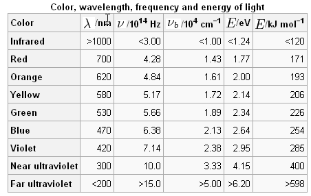
The wavelength
of the ultra weak photon emission (several million times weaker than
Bioluminescence) is measured in nanometres, which are very small, a thousand
millionth of a metre. A nanometre is notated as follows; 10−9
nanometre nm=0.000 000 001. Typical human eye will respond to wavelengths from
400 to 700 nm, although some people may be able to perceive wavelengths from
380 to 780 nm. Some of the research [35]
on ultra weak photon emissions is reporting UPE from 420 to 570 nm (Popp
reports 260 to 800 nm) [36]
with a range from 1 to 1,000 photons (x s-1 x cm-2)(I think this means photons
per second per square centimeter of surface area) [37].
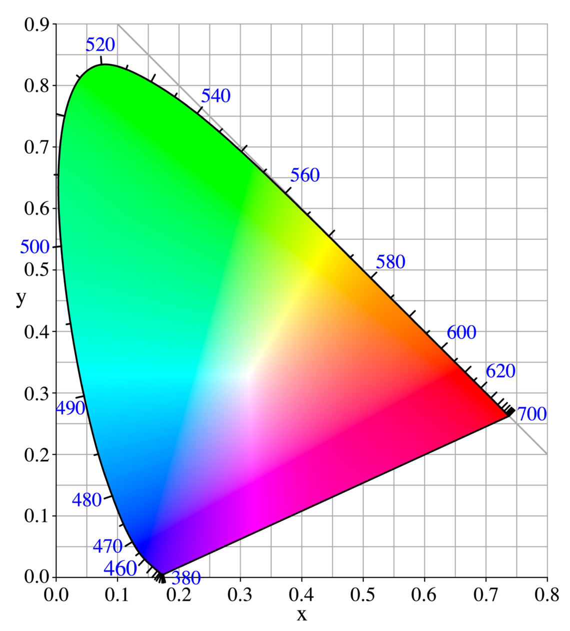
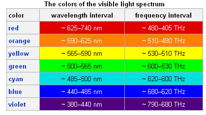
This range would correspond to the visible light color ranges of indigo
(Violet), blue, cyan, green and yellow (colors) (colors2)(colors3)
The wavelength is longer than greatest particle size that can fit through a
surgical mask but smaller than width of strand of spider web. Due to the low
concentration of photons it is not believed that these photons emissions can be
seen by the naked eye ("much weaker than in the openly visible" [38])
as in bioluminescence
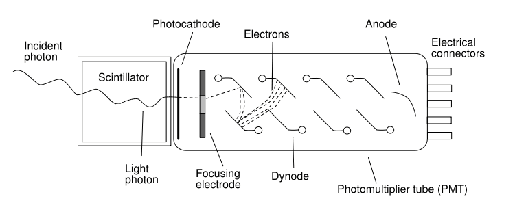
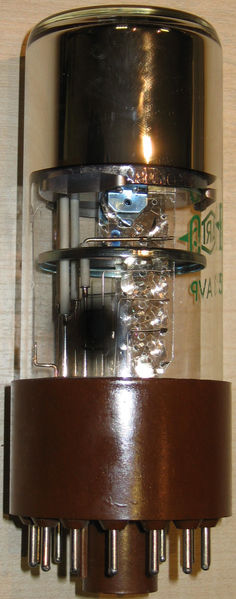
Photomultiplier
The detection of
Biophotons is facilitated by Photomultipliers (Photomultiplier tubes-PMTs) (Biophoton Tube Schematics)(Biophoton Tube)
which greatly amplify photons emitted in the ultraviolet, visible and near
infrared ranges. Photomultipliers are widely used in many fields (nuclear and
particle physics, astronomy, medical imaging and motion picture film scanning
(telecine) [39].
I could find no references of the use of photomultipliers however in the area
of medical imaging [40].
This is probably because this particular field of study is suspect. The
photomultiplier makes use of the photoelectric effect [41]
where photons hit a metallic surface and electrons are emitted. The
photomultiplier contains various electron capture devices (glass vacuum tube
which houses a photocathode, several dynodes, and an anode), which result in
the accumulation of charge and in a sharp current pulse indicating the arrival
of a photon at the photocathode. This device then can count the number of
individual photons produced from a variety of sources but for our purposes from
biological organisms.
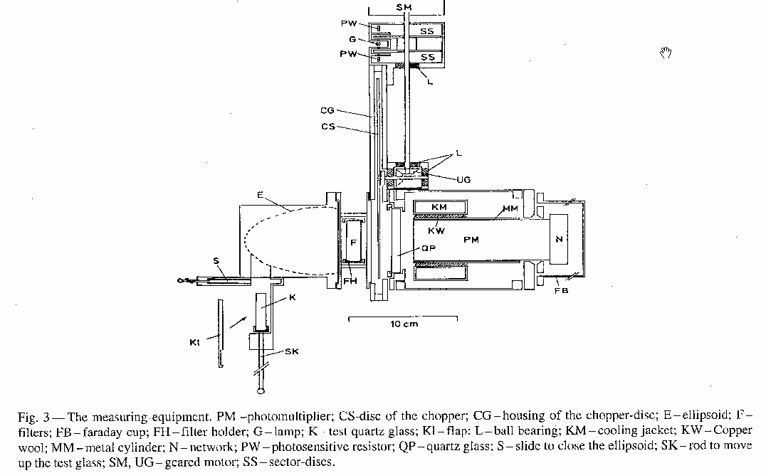
Popp describes
the photomultiplier that he uses as an EMI 9558 QA. Popp summarizes the
specifications [42]
from a more detailed dissertation paper [43]
as follows; This photomultiplier
uses a uses a "single photon counting system" with a sensitivity of
1017 W. 10 is the signal-to-noise ratio and the cathode has a range
sensitivity of between 200 to 800 nm. To reduce the noise to a minimum a copper
wool-cooling jacket "provides thermal contact". “A grounding metal
cylinder” accomplishes electric and magnetic field protection. The multiplier
tube and cooling jacket are housed in a vacuum and therefore the quartz glass
anterior to the tube in not in thermal contact with the cooled cathode thus
preventing moisture accumulation on its surface (resulting in freezing). With
this arrangement the optimal cooling temperature -30º C (Centigrade)(-22º F
Fahrenheit). A chopper (photomultiplier)
enhances current density to 2 photons/(s cm2) with a significance level of
99.9% within 6 hours.
Theoretical
Model-Biophoton Production-Mainstream Biophysicists
Although no
experimental proof for any definitive theory has been accepted even among the
field of experts, Biophotons are thought, by many biophysicists, to be random
photon emissions as a result of cellular metabolism. Given the 15 trillion
cells in the average human body (100 million in the brain alone), with the
average cell diameter of 10 micrometers, and the average photon emission of
1-1000 photons per second per square centimeter of surface area, this amounts
to a single photon per cell per month. Since cellular metabolism [44]
is a stepwise chain of small energy exchanges, occasionally mistakes are made
(random irregular steps (‘outlying states”)), which result in a physiochemical
energy imbalances and the rare emission of a photon. In other words it is the
occasional sour note in the symphony and not some orchestrated background
chorus.
According to
this hypothesis there is no need to attribute order where none exists, as does
the mitogenetic radiation hypothesis (see above). These
physiochemical energy imbalances occur as part of the electron transport chain
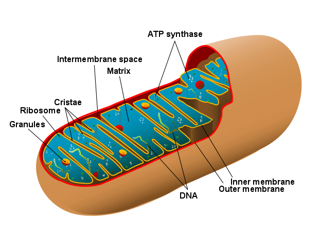
within the mitochondria
(Organelle) [45],
which is in every cell of the body. The electron transport chain creates
stepwise chemical reactions with the ultimate aim of creating useable energy
for cell metabolism. The mitochondria are known as the "cellular power
plants" because they convert organic materials into energy in the form of
ATP via the process of oxidative phosphorylation [46].
There are hundreds of thousands of mitochondria in every cell (can occupy 25%
of the cells cytoplasm)(mitochondria have their own DNA and may have once been independent
bacteria many millions of years ago). There are 105=100,000 or one
hundred thousand chemical reactions per cell/per sec and as aforementioned 15
trillion cells in the average human body (100 million in the human brain). We
are buzzing with activity. One purpose of the mitochondria is to create energy
for the cell to produce protein ect. Free Radicals (Reactive oxygen species or
ROS (superoxide, hydrogen peroxide, and hydroxyl radical)) are produced inside
the mitochondria and are associated with cell damage. Free radicals may be
created as a part of the production of ATP from ADP and may also be responsible
for the emission of Biophotons. The mitochondria produce energy
by converting ADP (Adenosine diphosphate) [47]
to ATP (Adenosine triphosphate) [48]
in a stepwise process along a protein matrix and the inner mitochondrial
membranes. The third step (electron transport chain) in this process involves
reattaching the phosphate group to ADP (Adenosine diphosphate) to form ATP
(Adenosine triphosphate). Once this is accomplished the cell can convert ATP
back into ADP and an inorganic phosphate producing the following amount of
energy; (12 kcal / mole in vivo (inside of a living cell) and -7.3 kcal / mole
in vitro (in laboratory conditions)). This third step as aforementioned is
called the electron transport chain in which electrons are stepped down in
energy by passing through a series of proteins. This way the lowered energy of
the electron can be safely utilized by the mitochondria. The third protein in
the electron transport chain is actually a lipid [49]
called Coenzyme Q [50].
Unfortunately 1-4% of the electrons that pass through Coenzyme Q leaks onto an
oxygen molecule in its outer shell (Open Shell configuration). This oxygen
molecule is called superoxide (O2) but it is unstable because it needs an
additional electron on its outer shell. Remember Coenzyme Q leaked an electron
onto its outer shell. Superoxide is prone to steal an electron from the nearest
source as follows; 1.) Mitochondrial DNA 2.) Mitochondrial Membrane (called
lipid peroxidation) 3.) Protein 4.) Reductants (Vitamin C, E, Non-Enzymatic
antioxidants (glutathione or thioredoxin). Borrowing electrons from Reductants
and Non-Enzymatic antioxidants does no harm to the cell. This is why you would
want to eat your vegetables and fruits because they contain antioxidants, which
lend electrons to the superoxide molecule which won’t then borrow from
structures such as mitochondrial DNA ect. Otherwise cell damage can result in
apoptosis, or programmed cell death. Not good for you.
According to
radical chemistry programmed cell death occurs as follows; “Bcl-2 proteins are
layered on the surface of the mitochondria, detect damage, and activate a class
of proteins called Bax, which punch holes in the mitochondrial membrane,
causing cytochrome C to leak out. This cytochrome C binds to Apaf-1, or
apoptotic protease activating factor-1, which is free-floating in the cell’s
cytoplasm. Using energy from the ATPs in the mitochondrion, the Apaf-1 and
cytochrome C bind together to form apoptosomes. The apoptosomes binds to and
activates caspase-9, another free-floating protein. The caspase-9 then cleaves
the proteins of the mitochondrial membrane, causing it to break down and start a
chain reaction of protein denaturation and eventually phagocytosis of the
cell.” [51]
The Free Radical
Theory of Aging [52]
advocates the use of antioxidants because they donate an electron to superoxide
without becoming unstable themselves. Aging occurs as mitochondria (cellular
power plant) become less functional or die out. As the cell can no longer
function and fail, aging accelerates. Free radicals like superoxide are an
inevitable by product of cellular metabolism but their damaging effects are
mitigated through the intake of antioxidants.
When Superoxide
borrows an electron from another source the theory is that a photon is
produced. This may be the explanation for ultraweak photon emissions. Since
this electron leakage only occurs in a small percentage of electron transfers
through Coenzyme Q the relatively low rate of photon emissions may be
consistent with this finding.
Theoretical Model-Popp
& Others-The Proponents
Biophotons are
involved in various cell functions, which include as aforementioned by
Gurwitsch cell mitosis and according to Russian, German, and other Biophotonics
experts may be produced and detected by the DNA in the cell’s nucleus.
Gurwitsch’s basic experiment ("Grundversuch") was the first example
of a proof that cell mitosis could be increased by UV (260 nm) alone after
carefully separating the inductor and detector plants with both a space of air
and alternately UV transparent and opaque glass. As whacky a proposition as
this is, the mostly vague dismissals by the mainstream biophysics community
will not dilute the implications. If replicated under strict controls
inevitable conclusions will demand explanation over extended time. The usual
Cell signaling mechanisms such as Notch signaling require physical contact
between the cells and or in the case of other cell to cell communication a
fluid medium such as blood (endocrine cells (Hormones)). Other cell-to-cell
communication is conducted thru interstitial fluid. Gurwitsch’s simple
experiment appears to thwart the usual mechanisms of cell signaling. The
conclusion is that Biophotons in the form of UV (260nm) emanating possibly from
the DNA of the inductor plant is signaling the DNA in the cell nucleus of the
detector plant to increase cell mitosis. Biophotons may then represent a more primitive
and yet subtly more complex cell-to-cell communication, which by passes the
usual fluid medium of information transmission and instead relies upon speed of
light transmission thru the air. (Does electromagnetic radiation within the
visible range transmit well through tissue? How and in what direction cell to
cell photon communication occurs between DNA strands may be unknown.
Gurwitsch was
himself an embryologist who was puzzling about how organs develop, and modern
Biophotonics experts suggest that Biophotons may offer some signaling mechanism
in the development of organs or other structures. Would electromagnetic carrier
waves such as radio, or light (fiber optics) inform us about the transmission
of information from cellular or mitochondrial DNA? Certainly before the
neurological or cardiovascular hardware was evolved electromagnetic
communication may have sufficed. Definitive proof is to date lacking. (?)
Given the 105=100,000
or one hundred thousand chemical reactions per cell/per sec, as aforementioned,
Popp states "Without electronic excitation of at least one of the reaction
partners, it would be impossible, and the number of thermal
photons in the tiny reaction volume of a cell could never suffice to explain
this high reaction rate. At least a 1014 (100,000,000,000,000=100
trillion) higher photon density in the optical range is necessary to provide
this huge amount of chemical reactivity." [53]
Given that not enough photons are produced in the cell there must be some other
explanation for the high chemical reaction rate within each and every cell.
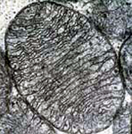
Erwin Rudolf Josef Alexander Schrödinger (1887–1961) may have led the way with
a simple observation and question. During cell division biomolecules must
migrate to either side of the cell as the two new cells form from one cell and
yet there are relatively few mistakes (“aberrations”) in this very complicated
process. Schrödinger simply asked his famous question why? A quick look at a
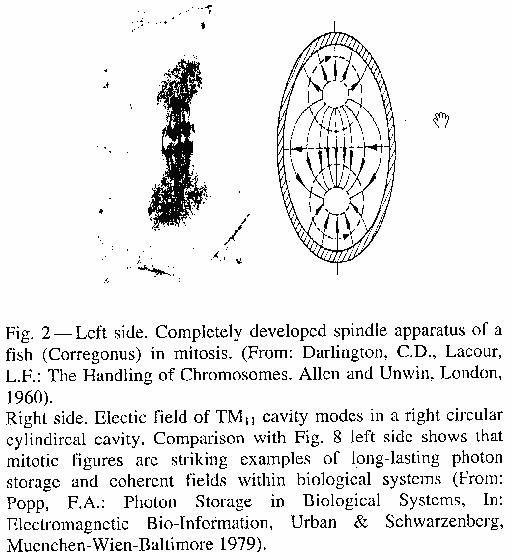
cell in mitosis on the left and an example of a cavity resonator wave on the
right is suggestive of an answer. Cell
Mitosis vs. Cavity Resonator Waves A cavity resonator wave in this
case is electromagnetic wave of a particular frequency (300-700nm) bouncing
back and forth between the walls of the cell, which somehow reflect these waves
with little loss of coherence. If more wave energy enters the cavity its
intensity is increased. This could explain the effects of Gurwitsch’s basic
experiment that by increasing to electromagnetic flow from the inductor plant
cell mitosis was increased in the detector plant. Popp believes that cavity
resonator waves are "the only plausible answer to this question" of
how there are relatively few mistakes during cell mitosis and with the
biochemical migration, which Popp thinks "also provide the necessary
stability of the molecular arrangements as the guiding forces for their
movement." [54]
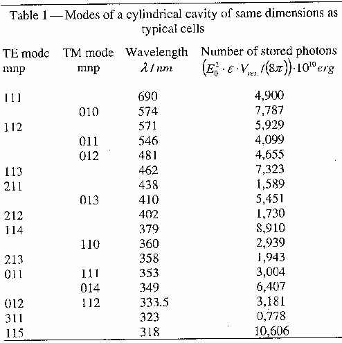
If the cell is viewed as a dielectric and or conducting resonant cavity, Popp,
demonstrates in Table 1 transverse magnetic
and electric modes and their wavelengths given the dimensions and boundary of a
cell. By superimposing the cavity resonator wave patterns onto the
"dynamical structures of the mitotic figures during cell division, Popp
reasons is "the most likely answer to Schrödinger 's question of why the
error rate vanishes". Popp acknowledges that there is no workable way to
measure these quasi-standing light waves directly within the intracellular
space although a photomultiplier placed near living tissue can measure single
photons within the visible range, which are correlated (spatial and temporal)
to cell mitosis. The more cell growth the greater the photon emissions. Around
1970 Popp organized an interdisciplinary group (University of Marburg
physicists, physicians, and biologists) to study the optical properties of such
biomolecules as polycyclic hydrocarbons (derived chiefly from petroleum and
coal tar?). Carcinogenic activity and other biological efficacy were studied
drawing out some questions of causality. Do the biomolecules themselves produce
photon ("light") emission or does some type of "photon
field" "the regulator for the excitation of biological matter."
Which causes which, chicken and egg conundrum. Popp puzzled over this question
proposing to characterize nonclassical light as a form of information transfer
in biological systems. What are the experimental results that support this bold
claim that biophotons can actually have a regulating function in biochemical
reactions? What is the physical basis for this and what are the theoretical
implications?
What are the
properties of biophotons, which are well described by multiple independent
groups and replicated numerous times [55]
[56]
[57]?
1.
The phenomenon
of photon emission from biological systems is quantum physical (coming from the
subatomic field within the organism?). Since fewer 100 photons are present (on
the surface) within the investigation field the total intensity i from a few up
to some hundred photons/(s cm2) confirms the quantum physical nature
of photon emission.
2.
What about the
nature of the biophoton emissions? The spectral intensity i(v) does not peak
around definite frequencies v. The characteristic of the spectral distribution
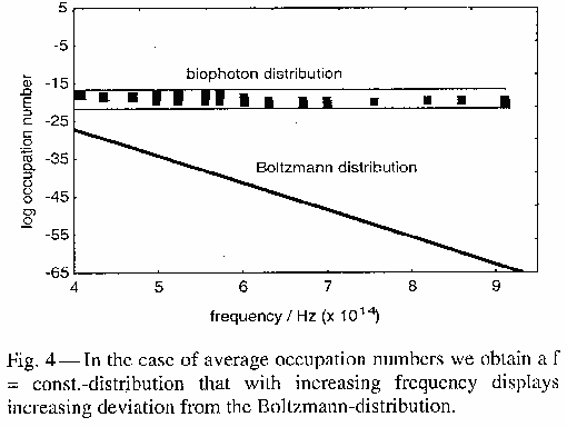
is flat and thus is a non-equilibrium system whose excitation temperature![]() (v) linearly increases, as does frequency v. The responsible excited
states of the occupation probability f(v) does not follow the Boltzmann
distribution f(v)=exp(-hv/kT) but the rule f(v)=constant (Fig. 4)
(v) linearly increases, as does frequency v. The responsible excited
states of the occupation probability f(v) does not follow the Boltzmann
distribution f(v)=exp(-hv/kT) but the rule f(v)=constant (Fig. 4)
3.
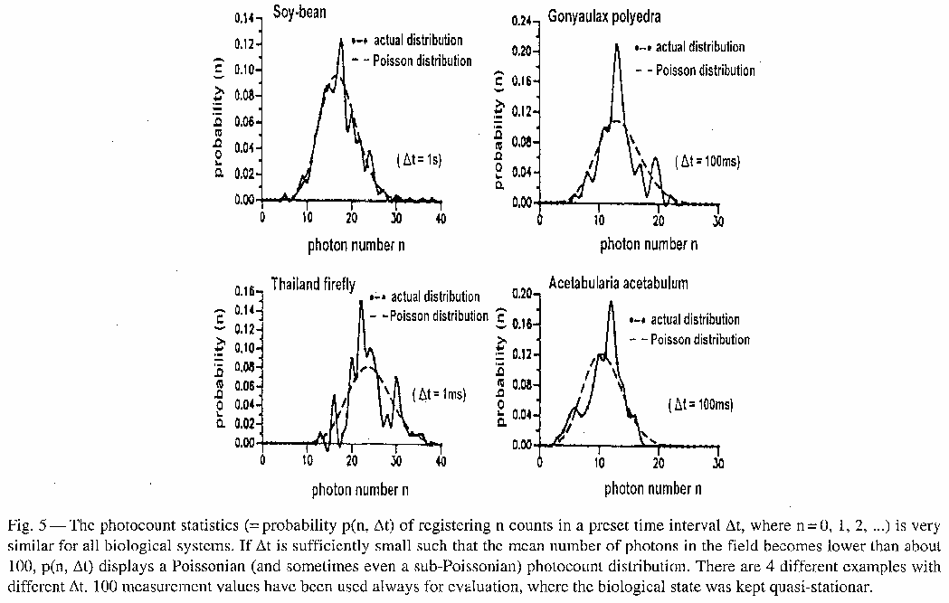
"The probability p(n, ![]() t) of registering n biophotons (n=0,1,2...) in a preset time
interval
t) of registering n biophotons (n=0,1,2...) in a preset time
interval ![]() t follows under ergodic
conditions surprisingly accurately a Poissonian distribution (exp(-<n>)
<n>n/n! <n>=mean value of n over
t follows under ergodic
conditions surprisingly accurately a Poissonian distribution (exp(-<n>)
<n>n/n! <n>=mean value of n over ![]() t time intervals
t time intervals![]() t down to 10-5
s. For lower time intervals t there are no results known up to now" [58] (Fig 5)
t down to 10-5
s. For lower time intervals t there are no results known up to now" [58] (Fig 5)
4.
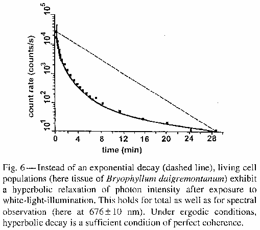
"Delayed
luminescence" (DL) (Long term and ultra weak reemission of photons after
exposure to monochromatic or white light illumination) diminishes with a
hyperbolic-like (l/t) function. Time after excitation=t There is no exponential
function evident in the diminution of photon emission. (Fig 6)
5.
The optical
extinction coefficient (fraction of light lost to scattering and absorption per
unit distance in a participating medium ) of Biophotons that penetrate thin
layers of sea sand and Soya cells (various thickness) was one order of
magnitude lower than artificial light tested in the same manner. The light
sources (biophotons/artificial) were matched for intensity and spectral
distribution and thus cannot be cited to explain the difference. Biophotons
loose less light when penetrating these mediums.
6.
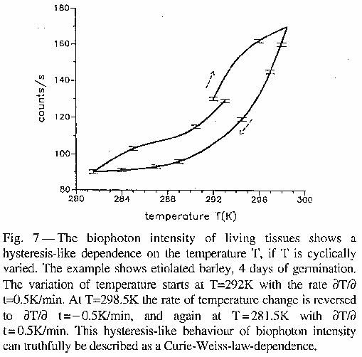
Physiological
functions such as membrane permeability and (Glycolysis) are known to be
affected by temperature and biophoton emission displays similar temperature
dependence. When temperature fluctuations occur both overshoot and undershoot
reactions occur. That is temperature increases cause overshoot and temperature
decreases cause undershoot biophoton emission reactions. These biophoton
emission fluctuation can be characterized as "temperature hysteresis
loops" (Fig
7) as described by a Curie-Weiss
law.
7.
As stress levels
increase so do biophoton emissions.
8.
Ethidium bromide
(EB) increases the unwinding (Conformation)
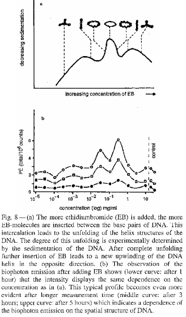
of DNA. Biophoton emissions are strongly correlated to the unwinding of DNA so
that when EB is intercalated into the DNA an increase in biophoton emission is
noted. (Figure 8)
This and other results suggests to Popp "that Chromatin (chromatino?)
is one of the most essential sources of biophoton emission. [59]
[60]
9.
Popp maintains,
"Biophotons originate from a coherent field". Evidence for this is
demonstrated in photocount statistics, which produce a Poissonian distribution.
These "photocount statistics p(n, ![]() t) under ergodic conditions together with hyperbolic relaxation function of
delayed luminescence is a sufficient condition of a fully coherent photon
field." [61]
t) under ergodic conditions together with hyperbolic relaxation function of
delayed luminescence is a sufficient condition of a fully coherent photon
field." [61]
There are biological phenomena, which can't be understood by
molecular biology or conventional biological thinking, which are better
explained if we assume biophotons originate from a coherent field. These
biological phenomena are better understood and predicted by biophoton theory.
The end result is a deepening of our collective biological understanding.
1.
Since the sum of
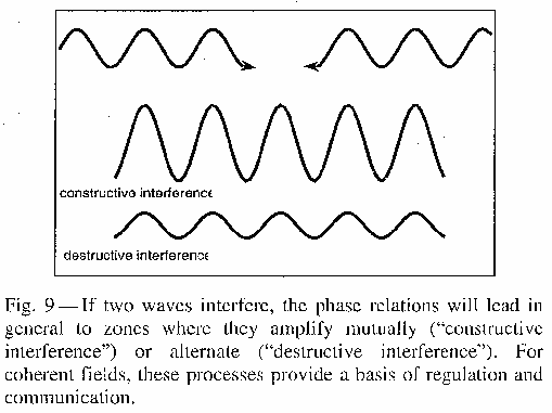
the energy has to remain constant in a closed system (Energy conservation law)
constructive interference (super-radiance) destructive interference (sub-radiance)
(Interference
Constructive and
destructive interference) serves the function of equity manager. (interference
example) (Fig.
9) (interference
example 2) Patterns of radiation according to Dicke [62]
are affected by time periods of interaction "between radiation and
non-randomly oriented matter of suitable size." Constructive interference
dominates in the initial interaction time period and destructive interference
dominates after longer time periods. Popp concludes that the probability of
destructive interference in intercellular space between living cells is high
for biophoton emissions.
2.
Since biophoton
fields between cells or living cellular organism cause interference patterns,
biophoton intensity (biophoton emissions counts?) is reduced. The emission from
single cells cannot be added up to find the total emission intensity because
biophotons are being canceled out by these interference patterns. Popp states
"biophoton intensity of living matter cannot increase linearly with the
number of units, but has to follow the effective amplitudes of the interference
patterns of the biophoton field between living systems." [63]
3.
The measurements
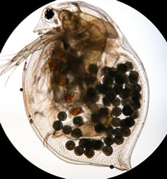
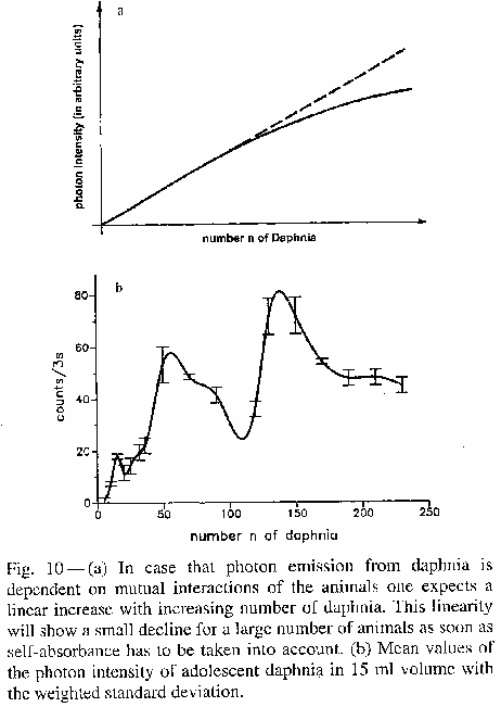
of biophoton emission of the planktonic, crustacean Daphnia magna [64]
[65]
illustrate the concepts explained in # 2 above. (Daphnia Magna Illus) The
biophoton emissions of these animals was measured under controlled conditions;
Darkness, housed within the quartz curvette of the biophoton measuring
equipment, Constant temperature 18º C (64.4º F). The numbers n (Independent Variable)
of daphnia was altered (1-250) maintaining equal size for these inbred animals.
The biophoton emissions (Dependent
Variable) were then measured after each increase in the count. Each
of these creatures emits about the same intensity of biophoton emission, which
means an increase in the number of animals should result in a linear increase
in biophoton emissions. Correcting for the self-absorption of biophoton
emission of the individual animal the biophoton emissions should look like the
linear graph in fig. 10 A (Fig.
10). Instead what was observed was the graph plot in Fig. 10 B. Popp
concludes "there is a tendency for destructive interference resulting in a
lower intensity than expected from the linear increase." [66]
In nature daphnia is found in concentrations of about 110 (Popp doesn't say per
square what?) animals. In this experiment at the same concentration (110) is
also creates the most efficient destruction zones around the organism (?) which
conserve stored light most effectively within the animals. The destruction zone
traps light within the animal according to the energy conservation law but as
aforementioned most efficiently at the natural concentration of 110 creatures.
4.
Popp states
"to some extent one is justified in saying that living systems
"suck" the light away in order to establish the most sensitive
platform of communication." [67]
[68]
Individual animals can be distinguished by similar wave patterns (?1), which are distinct
among species. Mutual interference patterns among groups of animals are also
distinct among species, which provides "necessary information about the
equality or difference of species." This mutual interference is a form of
biological communication. Each of the individual animals becomes aware of the
other thru biophoton communication (?2). The Signal-to-noise ratio (Signal Noise)
or mutual interference patterns (?3) are optimized at a
certain number of animals, which is also unique between species (?4). This optimization
results as aforementioned allows maximum light storage. (?5) This optimization is
achieved as noted by wave patterns, which interfere under maximum destruction
between the communication systems. Popp notes "every perturbation leads
then to an increase (signal) that the connected systems have to become aware
of." "This rather ingenious means of biocommunication provides the
basis for orientation, swarming, formation, growth, differentiation, and "gestaltbildung"
(?6)
in every biological system.” [69]
[70]
5.
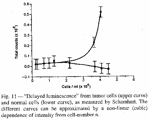
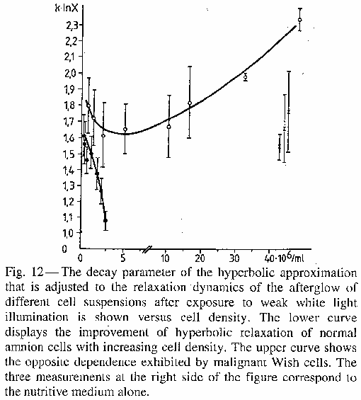
Damaged and or
destroyed tissue (first stage) (?1) [71]
affects the intensity of biophoton emission. The "capacity for coherent superposition
of modes of
the biophoton field (where longer wavelengths may also be included) breaks
down." As a result there is an increase in biophoton emission and or
delayed luminescence reflecting the breakdown of interference patterns between
the individual cells, which prevented the outward radiation of photon emission.
(?2)
[72]
Schamhart and Van Wijk [73]
(Fig. 11) [74]
(?3)
and Scholz et al. [75]
(Fig. 12) [76]
(?4)
were among the first to confirm this. Individual tumor cells, for example, loss
of coherence results in concomitant loss of destructive interference capacity
and delayed luminescence (converts from hyperbolic-like relaxation of normal
cells to exponential one of tumor cells).
6.
Dinoflagellates
exhibit asynchronous bioluminescence flickering when optically separated but
the opposite synchronous flickering when in optical contact. (Fig.13) [77]
When seen by other Dinoflagellates their bioluminescent flickering also
decreases. [78]
Bioluminescent is "chemically amplified biophoton emission" according
to Popp. The phenomena of destructive interference are, according to Popp, responsible
for flickering decreases and synchronous light pulses. "As the animals see
each other and displaying synchronous pulses as a consequence of the disruption
of the destructive interference patterns."
7.
Bacteria also
exhibit the same kind of communication within their nutrition media. [79]
8.
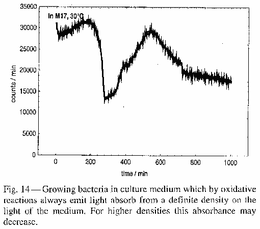
Fig. 14
illustrates the phenomena of bacteria (Enterococcus Faecalis)
grown in a nutrition media. The nutrition media emits biophotons as a result of
the oxygenation
processes. Therefore the nutrition medium produces a higher intensity of photon
emissions than the growing bacteria, which emit low biophoton intensity. Thus
the biophoton emissions of the bacteria are not registered. As the bacteria
grow in numbers their photon emission creates destructive interference within
the coherence volume of the light-emitting nutrient molecules. This results in
a drop in emitted biophotons at a specific bacteria number. As the number of bacteria
increase (Fig.
14) biophotons may again increase, as photons are no longer absorbed
thru destructive interference.
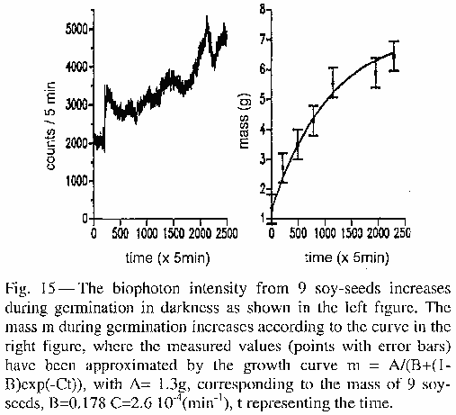
9.
Growth
regulation of biophoton emission follows the reciprocal laws where in addition
to linear stimulation n![]() n
(Proportionality
(mathematics)) nonlinear inhibition n
n
(Proportionality
(mathematics)) nonlinear inhibition n![]() n2
occurs in concert. That is to say there is a correlation between growth
rate and biophoton emission and that relationship is proportional as
aforementioned confirmed in Fig.
15.
n2
occurs in concert. That is to say there is a correlation between growth
rate and biophoton emission and that relationship is proportional as
aforementioned confirmed in Fig.
15.
10.
The presumptions
of Bajpai [80],
Gu and Li [81]
that organisms emit squeezed
light [82] as
opposed to classical coherent
light. These presumptions underlie a theoretical basis for biophoton
emission.
What theoretical perspectives can be derived from the
experimental observations outlined above and subsequently summarized? Certainly
classical electrodynamics
and thermodynamics
as well as quantum theory
provide a basis for biophoton theory. Biophoton theory as aforementioned will
need to explain the following summarized experimental results namely: spectral intensity
[83]
[84],
Photocount
statistics [85],
hyperbolic
oscillations [86]
[87],
coupling of the
different modes [88],
squeezing into both
branches of minimum uncertainty wave packets [89],
strong correlation
to DNA dynamical states [90].
Biological phenomena will also need explanation; mitotic figures (?) [91],
interference
structure from daphnia [92],
tumor tissue photon
emission vs. normal tissue [93],
and the correlation to growth
and differentiation of cells [94].
1.
(?1) Popp proposes an
equation to estimate the mean value of photons within a homogeneous
electromagnetic field. The Mean value is of the N=number of photons of
hv=Energy of a homogeneous electromagnetic field (?) with E0=Amplitude.
This mean value can be estimated by equating the energies nhv of the photons
and ![]() 0/(8
0/(8![]() ) | E0|2
V of the field where
) | E0|2
V of the field where ![]() 0=Dielectric constant and V=Volume of field. A
photon in the optical range of 3 eV equals a field amplitude of 106
V/cm over a cell volume of 109cm3. The aforementioned
draws the following conclusion; "Electric Field Amplitudes (of the cavity
modes) which stabilize the mitotic figures are in the range of 106
V/cm (corresponding to about the membrane field components). It would take only
one photon in the optical range would suffice for this effect." [95]
The ultraweak photon emissions can then be explained to reflect the requirement
of only one photon to provide for the biological functions within the cells
which include; "stabilization of the migration of the biomolecules,
transportation of the angular momentum for rotating the DNA during replication
or transcription, and provision of the chemical reactivity of about 105
reactions per cell and per second, always at the right time and at the right
place." [96]
0=Dielectric constant and V=Volume of field. A
photon in the optical range of 3 eV equals a field amplitude of 106
V/cm over a cell volume of 109cm3. The aforementioned
draws the following conclusion; "Electric Field Amplitudes (of the cavity
modes) which stabilize the mitotic figures are in the range of 106
V/cm (corresponding to about the membrane field components). It would take only
one photon in the optical range would suffice for this effect." [95]
The ultraweak photon emissions can then be explained to reflect the requirement
of only one photon to provide for the biological functions within the cells
which include; "stabilization of the migration of the biomolecules,
transportation of the angular momentum for rotating the DNA during replication
or transcription, and provision of the chemical reactivity of about 105
reactions per cell and per second, always at the right time and at the right
place." [96]
2.
Popp states, "living systems may be
looked upon as the most stable forms of matter through use of the storage of
sunrays" with the resonators model as a powerful tool in understanding
biophoton emission. [97]
Since the sun is very hot and the earth is comparatively very cold the light
from the sun either reflects from the earth or through a process of entropy
transforms heat to cold. To sustain life organisms must prolong this process by
optimizing their "storage capacity for sunlight." [98]
In plants for example photosynthesis
provides for its elementary food supply by synthesizing glucose from sunlight,
carbon dioxide, and water. Animals get their glucose from plants, protein from
other animals and as Popp proposes uses sunlight to guide the molecular biology
of the cell and literally spark biomolecular processes to convert ADP![]() ATP to provide the
energy for cellular metabolism.
ATP to provide the
energy for cellular metabolism.
3.
A resonator
value within any cavity (including the cell) can be determined and Popp states
that there is a "clear connection between the resonator value of a cavity
and its information content." This establishes some key understandings of
biological systems namely; These biological systems are informational rather
than energetic "engines", and the resonators may develop nonlinear
capacities "just because of their low photon emission." [99]
The equation, (Q*=Q/1-C), represents the deviation from the classical Q-value (Q factor (Q-Value)) of the
typical resonator. The variables are defined as follows; Q*=resonator value of
the quantum coherent resonator Q=value of the classical "chaotic"
resonator C=ratio of a quantum coherent energy distribution of the resonator to
the totally available (chaotic + coherent) energy.
4.
The high storage
time and ability to emit or to remove photons actively for C>1 is reflected
in this equation; Eq. # 1=(Q*/Q![]()
![]() for C
for C![]() 1). Bose-Einstein
condensate (Bose-condensation) (Bose-condensation-like phenomena) as
postulated by Herbert
Fröhlich can also be explained by taking the Bose–Einstein statistics
(Bose-Einstein Distribution) "of the spectral photon density
(number of photons per units of volume and wavelength
1). Bose-Einstein
condensate (Bose-condensation) (Bose-condensation-like phenomena) as
postulated by Herbert
Fröhlich can also be explained by taking the Bose–Einstein statistics
(Bose-Einstein Distribution) "of the spectral photon density
(number of photons per units of volume and wavelength ![]() ) at temperature Eq.
# 2=TN(
) at temperature Eq.
# 2=TN(![]() )=8
)=8![]() /
/![]() 4 1/(exp((
4 1/(exp((![]() -
-![]() )/(kT))-1) where
)/(kT))-1) where ![]() =hc/
=hc/![]() is the photon energy and
is the photon energy and ![]() the chemical
potential, and k is the Boltzmann
constant.” [100]
the chemical
potential, and k is the Boltzmann
constant.” [100]
5.
The chemical
potential is defined as ![]() =T(
=T(![]() (?)S/
(?)S/![]() n)e,v where dS is the entropy change through absorption of a
photon. Entropy in the system is increased along with the value of
n)e,v where dS is the entropy change through absorption of a
photon. Entropy in the system is increased along with the value of ![]() >0 when a biophoton is absorbed by the multiplier outside
the system (dn<0) as depicted in Fig.
2. When there is no entropy loss due to thermal noise then
>0 when a biophoton is absorbed by the multiplier outside
the system (dn<0) as depicted in Fig.
2. When there is no entropy loss due to thermal noise then ![]() =
=![]() . Also possible is Eq.
# 3=
. Also possible is Eq.
# 3=![]() =
=![]() -kTlnW where W corresponds to the thermo dynamical
probability of the photons under investigation. Insertion into Eq. (2) results
in Eq. # 4=N(
-kTlnW where W corresponds to the thermo dynamical
probability of the photons under investigation. Insertion into Eq. (2) results
in Eq. # 4=N(![]() )=8
)=8 ![]() /
/![]() 4 1/(W-1).
This demonstrates the Bose-Einstein
condensate (Bose-condensation) (Bose condensation effect) of the
Fröhlich mode (?) according to W
4 1/(W-1).
This demonstrates the Bose-Einstein
condensate (Bose-condensation) (Bose condensation effect) of the
Fröhlich mode (?) according to W![]() 1 as well as the
connection to the corresponding value C in Eq. (1). C=1 determines that all of
the energy of this system conforms to a coherent field except the classic
currents. In the case of classical currents resonance-like absorption of
photons in the mode W
1 as well as the
connection to the corresponding value C in Eq. (1). C=1 determines that all of
the energy of this system conforms to a coherent field except the classic
currents. In the case of classical currents resonance-like absorption of
photons in the mode W![]() 1 occurs.
"Squeezed" light would describe removal of photons by W>1 or the
extension of W, where the thermo dynamical potency of the photon field corresponds
to the vanishing chemical potential according to Eq. # 3. Eq. # 5=Ln W=
1 occurs.
"Squeezed" light would describe removal of photons by W>1 or the
extension of W, where the thermo dynamical potency of the photon field corresponds
to the vanishing chemical potential according to Eq. # 3. Eq. # 5=Ln W=![]() /(kT); This results in a spectral intensity of thermal
radiation. Can we determine the nature of biophoton emission by analyzing its
average spectral intensity? W turns out to be rather constant and indepencent
of the wavelength (see Fig. 16=There was no figure 16 in the research article I
had)
/(kT); This results in a spectral intensity of thermal
radiation. Can we determine the nature of biophoton emission by analyzing its
average spectral intensity? W turns out to be rather constant and indepencent
of the wavelength (see Fig. 16=There was no figure 16 in the research article I
had)
A significant increase in photon emission is evident around
sites of tissue injury, as do injured organisms prompting some Biophotonics
experts to suggest this could be a “distress signal” possibly to promote wound
healing. The mainstream critics are quick to remind that cellular damage
increases oxidative stress, electron leakage, and increased concentration of
superoxide with the greater potential for electron swapping and thus increased
photon production. However proponents argue a correlation between greater wound
healing and increased photon production, which reverses with lower levels of
photon emission. Could Biophoton emissions for example signal malignancy in
tissue before more conventional imaging? Do photons transmit thought just as
the nervous system? Even the experts answer these questions differently but the
answers cover the spectrum.
Perhaps, say proponents, Biophoton emissions are primitive
neural systems used by single celled organisms as they developed into more
complex creatures. Biophotonic signaling may also be used in modern complex
organisms, such as us, in the reception, transmission, and processing of
electromagnetic data perhaps with some of the same transmission features of
fiber optics or radio waves.
The Skeptics
The skeptics argue that mainstream biological sciences and
biophysics regard Biophotonics as pseudoscience [101],
which has been, relegated to the fringe; References in respectable journals are
virtually unknown. (Is this true?) According to doubters Signal noise or artifacts
from the measuring equipment (photomultipliers) as aforementioned are
responsible for photon production and represent random noise and no coherent
cell-to-cell communication. This phenomena of Biophotons although as all agree
is a natural phenomena has no meaning beyond that. Just as the bumps in a
persons cranium does not reveal traits of personally (Phrenology) or the
conjunction of the planets predict wars (Astrology). We as humans are
pattern-seeking creatures, which may relate to the evolutionary need to avoid
being eaten (establish and predict the movements of friend or foe both with
movements and coloration for example). This natural pattern seeking affects
even scientists who observe patterns in nature which in fact does not exist, or
marketers for example who create colorful logos and marketing campaigns to get
us to buy product.
Cell to cell communication is further compromised by the
relatively more intense sunlight or even starlight, which would interfere with
any photon signaling. Conversely proponents argue that this "kind of
signaling involving entangled quanta of light (e.g. Biophotons) can't be
swamped out by classical light, the same way a laser beam can still send
information in bright daylight, coherence affords "special privileges."
AW
New age, complementary and alternative medicine, and quantum mysticism profit
mongers are selling Biophotons in health cures for serious illness such as
cancer causing people to postpone more effective but conventional treatments.
In short this is metaphysics and not science. The field of Biophotons is rife
with new age devises and Web sites which promote the Biophotonics proof that
healing energy exists and can restore health.
Web Sites
http://www.google.com/search?hl=en&q=biophoton+healing
Quantum Mysticism
Fugue-Implications of we are light (substitute light for energy where
appropriate)
Taoist cosmology (secrete teachings)(I am using broad
strokes-its been many years and some factual errors may exist) believes that
over many lifetimes we give birth to our energy body, which is housed, in our
abdomen in what is known as the crystal palace. It is between the navel and 3rd
lumbar vertebrae. This energy body can be used by our spiritual consciousness
(housed between the pineal and third eye point between the eyebrows) to break
free from the cycles of birth and death. When we die for example our
consciousness, whose signature is embedded in light, continues without a
physical body but without an energy body is unstable and longs to return to
physicality. Our task in the physical realm is to give birth and mature an
energy body, which can sustain spiritual consciousness, the energy body can
only grow and mature in the physical realm (Taoist masters may disagree on this
point) , that is when we have a physical body. In order to do this the energy
body must be nurtured in a neutral energy environment. Strong emotions for
example must be transmuted into this neutral energy. This includes too much
anger or kindness, fear or gentleness, joy and spitefulness ect. This is why
Taoism is about a balanced path. Each organ houses the excesses of these emotions.
All of this is carried by our consciousness after physical death imprinted and
stored in a light signature. This is likened to the slow process of creating a
pearl only at the center of the pearl is the energy body and the outer layers
are made of light energy. Both the energy body and consciousness are poised
between two large energy balls above and below our heads (The Indian system I
believe calls these atman and Brahman.) Disease can be as an imbalance of
energy streaming between these balls and stored overly positive or negative
emotions in the organs and channel system. The energy channels are composed of
light waves, which transmit this energy. Once an energy body is stabilized we
become enlightened beings similar to Abraham Maslow’s (1908–1970) actualized
being. This is also reminiscent of Mesmer’s theory of Animal Magnetism. Perhaps
our spirit is a light wave signature embedded in Eugenio Calabi’s
(1923-present) and Shing-Tung Yau’s (1949-present) Calabi-Yau manifold.
9219/300= 30.73
Notes
10−9 nanometre nm
420-470 nm 470-570 nm
range from 1 to 1,000 photons x s-1 x cm-2
420-440 nm — wavelength of indigo light
440-500 nm — wavelength of blue light
500-520 nm — wavelength of cyan light
520-565 nm — wavelength of green light
565-590 nm — wavelength of yellow light
γ = Gamma rays
HX = Hard X-rays
SX = Soft X-Rays
EUV or XUV= Extreme ultraviolet (1–31 nm)
FUV or VUV=far or vacuum UV (200–10 nm)
NUV = (380–200 nm)
Near ultraviolet
Visible light
NIR = Near infrared (0.75–1.4 µm)
MIR = Moderate infrared
FIR = Far infrared
Radio waves:
EHF = Extremely high frequency (Microwaves)
SHF = Super high frequency (Microwaves)
UHF = Ultrahigh frequency
VHF = Very high frequency
HF = High frequency
MF = Medium frequency
LF = Low frequency
VLF = Very low frequency
VF = Voice frequency
ELF = Extremely low frequency
Need Definitions
Photon counting techniques, refractive index matching,
bioluminescence, biophotons, high quantum efficiencies, C2550 photon counter
(Hamamatsu Photonics K.K.), R647 (1/2 inch), R331 (2 inch),
and R329 (2 inch) photomultiplier tubes (PMT, Hamamatsu Photonics K.K.),
bialkali photocathode, spectral response, mode coupling, Steady State Biophoton
Emission, Poisssonian Photo Count Distribution, fully Coherent, Squeezed
States, Thermodynamic and Quantum Optical Interpretation,
Gestalthbildung=Swarming, Non-Thermal Photon Vs. Thermal Photons Emission, Cavity Resonator Waves,
long lasting photon storage, resonance wavelengths, transverse magnetic
and electric modes, dielectric resonant cavity, (eigenvalues of the Bessel
functions m, n correspond to the radial axis and p to the length of a right
circular cylindrical cavity.), TE mode mnp TM mode mnp, Number of stored
photons, superposition of cavity resonator waves, Schrödinger 's question of small number of
aberrations in the migration of biomolecules during cell division, polycyclic
hydrocarbons, (photon counting system functions at a sensitivity of about 1017
W and a signal-to-noise ratio of at least 10), EMI 9558 QA photomultiplier
cathode sensitive within the range of 200-800 nm,
(decay parameter, hyperbolic approximation, relaxation dynamics, cell
suspension afterglow, weak white light illumination, normal amnion cells, cell
density, malignant Wish cells, nutritive medium. (Fig.
12))
1.
What is a thermal photon
and how are the number of these counted within a single cell anyway? I thought
counting photons within a single cell was impossible? Is a thermal photon
different from other ultraweak photons under discussion? Reference; Popp Fa.
(2003). Properties of biophotons and their theoretical implications. Indian J
Exp Biol, 41-5, pp. 391 - 402. Full Text Article; http://www.anatomyfacts.com/research/PropertiesBioph.pdf
2.
Given that the
high chemical reaction rate 105=100,000 per cell per sec the number
of thermal photons says Popp are insufficient to explain this high reaction
rate? In other words you would need vastly more photons in the cell to explain
the high reaction rate on the order of 1014 (100,000,000,000,000=100
trillion) since at least one of the chemical reactants needs a little
electrical buzz to allow the chemical reaction. Popp implies that these are the
other biological phenomena (high number of chemical reactions), which explain
the existence of photons within cells. Is there a disconnect here? It doesn’t
seem to be explained well. Reference; Popp Fa. (2003). Properties of biophotons
and their theoretical implications. Indian J Exp Biol, 41-5, pp. 391 - 402.
Full Text Article; http://www.anatomyfacts.com/research/PropertiesBioph.pdf
3.
Is cell-to-cell
signaling an accepted scientific fact which explains "bloom of
bioluminescent algae creating an entrainment of light pulsing" AW. What is
the mechanism ect? Could the same mechanism be at work between the cells of
organisms only in this case ultra weak photon emissions and has this been
considered?
4.
What evidence do
we have on "cell-to-cell signaling within the human CNS through
biophotons" AW? The reference in this paper is CNS.
5.
Please expand on
the "implications (biophotons) this could have for developmental biology,
as well as injury healing." AW
6.
What is
“structured water”? AW
7.
What is “NAD,
and CoQ10”? AW
8.
What is the
"ubiquitous and critical process of gel/sol transition states in all
biological systems" AW?
9.
What is Coherent
non-classical light and optical coherence? Contrast and compare classical light
terms vs. non-classical light terms with regards to biophotons.
10. Talk about non-classical or squeezed light behaviors
referencing quantum entanglement aka Einstein’s " "spooky action at a
distance."
11. How can we measure single photon behaviors?
12. What is the experimental evidence supporting quantum
entanglement?
13. What is "sub-threshold" photon counting"?
14. What is spontaneous emission of photons via vacuum
fluctuations?
15. Perhaps then these quantum entanglement "spooky action
at a distance" are like some cosmic tug of war in the fabric of space,
where simultaneity does not violate special relativities speed limit (SOL). Its
like pulling on a rope at some summer back yard barbeque. Perhaps at the
cellular level we might observe similar effects. Are there cancers for example
that simultaneously appear in different parts of the body with no currently
known route of transmission? Do these weak photon emissions transmit data like
some fiber optics. How do radio waves or any electromagnetic waves transmit
data? Can data be stored and preserved?
16. What are the applied Biophysics books edited by Popp? More
information about summer school/conferences at Neuss?
17. Are these articles listed the best ones to do a literature
review on? http://www.anatomyfacts.com/Muscle/photonr.html
18. What is “Van Wijk's paper on Human Biophoton counting” AW?
19. Has Gurwitsch’s basic experiment ("Grundversuch")
been replicated?
20. How did Gurwitsch determine that particular range of UV light
(260nm) was being emitted?
21. By what process does the DNA of one plant produce DNA
signaling to another plant cell? Is this one way or two ways? Is this the same cell-to-cell
communication we see in animals? Have we been able to image any of this and if
so by what technology is imaging possible?
DNA plays a role in things, but not in the sense that is
normally thought. AW
22. Would injury to a cell for example increase photon production
from other healthy cells to stimulate the DNA of the injured cell to facilitate
healing?
23. Wouldn't the by many times multiplied relative intensity of
sunlight for example interfere with the ultra weak photon emission cell to cell
signaling?
The kind of signaling involving entangled quanta of light
(e.g. biophotons) can't be swamped out by classical light, the same way a laser
beam can still send information in bright daylight, coherence affords
"special privileges." AW We nee references for this. TN
24. Do we have an English translation of the full text version of
the paper that took us down this rabbit hole in the first place? A.G.
Gurwitsch: "Über Ursachen der Zellteilung". Arch. Entw. Mech. Org. 51
(1922), 383-415
25. Popp, provides us with a good historical review and then
seems to suggest that cell mitosis is guided by electromagnetic resonant waves
(?)(300-700nm), which would explain Erwin Schrödinger’s question regarding how
there could be so few errors in the biomolecular migration during cell mitosis.
It is in Table 1 that I become lost. What is the meaning of Table 1? Reference;
Popp Fa. (2003). Properties of biophotons and their theoretical implications.
Indian J Exp Biol, 41-5, pp. 391 - 402. Full Text Article; http://www.anatomyfacts.com/research/PropertiesBioph.pdf
26. Who is Dr. Sutherland and what is his experience with regards
to scientific skepticism?
William Garner Sutherland DO (1873-1954) was a student of Andrew
Stills (circa 1900) who believed the bony cranium was capable of respiratory
motion. "While looking at a disarticulated skull, Sutherland was struck by
the idea that the cranial sutures of the temporal bones where they meet the
sphenoid bones were "beveled, like the gills of a fish, indicating
articular mobility for a respiratory mechanism."” Dr. Sutherland "the
cranial-osteopath who laid the foundation for cranio-sacral therapy especially
the biodynamic branch. "Liquid
Light" he would say is the property of inherent health expressing within
the body." AW
27. What does BG stand for as used in the phrase “BG Chem and
physics”?
28. Can we determine whether or not the Taoist Cosmology,
traditional Eastern medicine and Channel theory has any merit? For example do
photon emissions seem to concentrate along the traditional channel lines such
as Stomach or liver channels in the leg or lung and large intestine channels in
the arms? Can we see photons produce an aura and is there any photon research
to show how energy work effects. Do we find increased photon emissions above
and below the head and below the feet for example? Is there a greater
concentration of photon emission around the crystal palace area or near the
third eye? Does more energy come out of the hands when energy work is being
done? Do energy workers produce a significant increase in cell mitosis in
plants when compared to Gurwitsch’s basic experiment? What statistical tools
should be employed?
29. If in Biophotonics human energy fields can be photographed,
what is the technology used?
30. Is our spirit is a light wave signature embedded in Eugenio
Calabi’s (1923-present) and Shing-Tung Yau’s (1949-present) Calabi-Yau
manifold?
31. How do the interior walls of a cell reflect electromagnetic
waves (Cavity resonator waves)? Do cavity resonator waves help guide
biochemicals and reduce error rate during cell mitosis?
32. Do cavity resonator waves explain the effects of Gurwitsch’s
basic experiment that by increasing the electromagnetic flow from the inductor
plant cell mitosis was increased in the detector plant?
33. Does the sweet reason Popp uses to justify cavity resonator
waves answering Schrödinger 's question a plausible explanation to other
biophysicists? Is there experimental proof of this. The reference is as follows;
Popp, demonstrates in Table 1 transverse magnetic
and electric modes and their wavelengths given the dimensions and boundary of a
cell. By superimposing the cavity resonator wave patterns onto the
"dynamical structures of the mitotic figures during cell division, Popp
reasons is "the most likely answer to Schrödinger 's question of why the
error rate vanishes".
34. What is the “electric field of TM11 cavity modes
in the Right side explanation of Fig. 2 in this illustration Cell
Mitosis vs. Cavity Resonator Waves?
35. What are the eigenvalues of the Bessel functions m, n in Table
1 and how do they correspond to the radial axis and p to the length
of a right circular cylindrical cavity? In the same table what is TE mode mnp
TM mode mnp and what’s the concept and actual number of stored photons mean?
36. What are the experimental results that support this bold
claim that biophotons can actually have a regulating function in biochemical
reactions? What is the physical basis for this and what are the theoretical
implications?
37. What are the single photon counting system functions? W=Wattage?
Signal-to-noise ratio. What’s a cathode? What kind of photomultiplier is the
EMI 9558 QA. What do range sensitivities mean (200 to 800nm)? Why does
inserting the multiplier into a cooling jacket, where copper wool provides
thermal contact, reduce the noise? How does a grounding metal cylinder protect
the multiplier from electric and magnetic fields? Why does freezing occur if
the multiplier is not kept in a vacuum? Why does the quartz glass in front of
the multiplier tube have no thermal contact with the cooled cathode? Why
doesn't it become covered with moisture? Why is the optimal cooling temperature
-30º C (Centigrade)(-22º F Fahrenheit)?
What is a chopper? What is current density (2 photons/(s cm2)?
What is significance level (99% within 6 hr.)?
38. What is quantum
physical (coming from the subatomic field within the organism?)?
39. There are only three references in this section and none of
them appear independent. What are the multiple independent groups and
replicated studies? (Studies?)
40. Please interpret the following # 2 (Boltzmann)
41. What is spectral intensity, non-equilibrium system,
excitation temperature![]() (v), occupation probability f(v), Boltzmann distribution
f(v)=exp(-hv/kT) but the rule f(v)=constant.
(v), occupation probability f(v), Boltzmann distribution
f(v)=exp(-hv/kT) but the rule f(v)=constant.
42. Please explain the following terms; probability
p(n, ![]() t) n biophotons (n=0,1,2...) preset time interval
t) n biophotons (n=0,1,2...) preset time interval ![]() t ergodic conditions Poissonian distribution (exp(-<n>)
<n>n/n! <n>=mean value of n over
t ergodic conditions Poissonian distribution (exp(-<n>)
<n>n/n! <n>=mean value of n over ![]() t time intervals
t time intervals![]() t down to 10-5
s
t down to 10-5
s
43. Please
interpret this statement "The probability p(n, ![]() t) of registering n biophotons (n=0,1,2...) in a preset time
interval
t) of registering n biophotons (n=0,1,2...) in a preset time
interval ![]() t follows under ergodic conditions surprisingly accurately a Poissonian
distribution (exp(-<n>) <n>n/n! <n>=mean value of
n over
t follows under ergodic conditions surprisingly accurately a Poissonian
distribution (exp(-<n>) <n>n/n! <n>=mean value of
n over ![]() t time intervals
t time intervals![]() t down to 10-5
s. For lower time intervals t there are no results known up to now" [102] (Fig 5)
t down to 10-5
s. For lower time intervals t there are no results known up to now" [102] (Fig 5)
44. What
is a hyperbolic-like (l/t) function? (Fig 6)
45. What is the optical
extinction coefficient?
46. Please explain how temperature
increases and decreases cause "temperature hysteresis loops" (Fig 7) as
described by a Curie-Weiss
law.
47.
Popp uses the
word chromatino is this the same as chromatin? http://www.anatomyfacts.com/research/PropertiesBioph.pdf
48. Are the DNA strands separated from other cellular material so
that the DNA can be the only source of biophoton emission?
49. Does the phenomena of destructive interference explain the
phenomena of ultra-weak photon emissions? (Interference reference)
Given the large role these electromagnetic waves have in all biological
functions wouldn’t you expect greater intensity photon emissions?
50. What does Popp mean by the statement "biophoton
intensity of living matter cannot increase linearly with the number of units,
but has to follow the effective amplitudes of the interference patterns of the
biophoton field between living systems."? [103]
(Reference)
51. Could the elimination of destructive interference explain
phenomena such as spontaneous combustion of living organisms? (Reference)
52. How do these destruction zones trap light within the organism
and does the same mechanism work intercellular? (Reference)
53. What is the daphnia concentration in nature that Popp
references? (Reference)
He found in concentrations of about 110 (Popp doesn't say per square what?)
animals. Can we get an English translated copy of the referenced dissertation
article? [104]
54. Is this energy that is trapped within the creatures from
sunlight? (Reference)
55. What does Popp mean by the statement "to some extent one
is justified in saying that living systems "suck" the light away in order
to establish the most sensitive platform of communication."? [105] Where does this light come from? (Reference)
56.
Can individual
animals be distinguished by similar wave patterns and if so could this be used
to identify certain bacteria within cultures or types of cancer tumors within
people? (Biological phenomena
4 Q1)
57.
Is this mutual
interference is a form of biological communication and how does it work? How
does each of the individual animals become aware of the other thru biophoton
communication? (Biological
phenomena 4 Q2)
58.
Is the
Signal-to-noise ratio the same as mutual interference patterns? (Biological phenomena 4 Q3)
59.
Is the number of
animals necessary for the optimization of the mutual interference pattern
unique to individual species? If it is, could bacteria for example be further
identified by the number of animals required for the optimization of the mutual
interference pattern? (Biological
phenomena 4 Q4)
60.
How does this
optimization allow for maximum light storage? What experimental proof do we
have? (Biological phenomena
4 Q5)
61.
How do biophoton
emissions provide the biocommunication necessary for orientation, swarming,
formation, growth, differentiation and "gestaltbildung”? What is swarming?
What is "gestaltbildung”? (Biological
phenomena 4 Q6)
Gestaltbildung
is the formation and differentiation of tissues and organs.
62.
What does Popp
mean by the “first stage” of tissue destruction? (Biological phenomena 5 Q1)
63.
Are these
interference patterns also responsible for maintaining coherence within
individual cells (Maintain “Cavity resonator waves” within cell)? Once tissue
destruction occurs and interference patterns are eliminated where do biophoton
emissions come from? Why does delayed luminescence increase exponentially
subsequent to tissue destruction and interference pattern elimination? How does
the breakdown of interference patterns between the individual cells occur from
tissue destruction? (Biological
phenomena 5 Q2)
64.
What is
non-linear (cubic) dependence of intensity from cell-number n in (Fig. 11)?
(Biological phenomena
5 Q3)
65.
Please rephrase the description
under (Fig. 12)
with "layperson friendly" definitions of the following terms; (decay
parameter, hyperbolic approximation, relaxation dynamics, cell suspension
afterglow, weak white light illumination, normal amnion cells, cell density,
malignant Wish cells, nutritive medium.) (Biological phenomena 5 Q5)
66.
Please make this
equation layperson friendly. (Theoretical
Perspective 1 Q1)
1.
Skeptical
dissertation of Biophotonics AW TN
2.
Synopsis of BG
physics and chem. AW
3.
Profile on the
typical reader of JBMT. TN
4.
Literature
Review Misc Articles AW TN
5.
Literature
Review Photon Counting for injured tissue AW TN
6.
Literature
Review of articles examine the implications of
“We are made of light” Taoist Cosmology, Eastern Medicine and Western
energy work AW TN
7.
Send lit review
to both skeptic and proponent biophysicists and others for review.
8.
E-Mail online
Chat list with identified interest area.
9.
Start local
Journal Club So. Ca. TN
Massage
Journal Club Online was initiated on 11/6/2006 http://health.groups.yahoo.com/group/journalclubonline/
Unit Presentations are scheduled during November to recruit interested members.
10.
Glossary
Basic aromatic ring
Basic aromatic rings are aromatic rings in which the lone
pair of electrons of a ring-nitrogen atom is not part of the aromatic system
and extends in the plane of the ring. This lone pair is responsible for the
basicity of these nitrogenous bases, similar to the nitrogen atom in amines. In
these compounds the nitrogen atom is not connected to a hydrogen atom. Basic
aromatic compounds get protonated and form aromatic cations (e.g. pyridinium)
under acidic conditions. Typical examples of basic aromatic rings are pyridine
or quinoline. Several rings contain basic as well as non-basic nitrogen atoms,
e.g. imidazole and purine.
Biophotonics
Popp’s definition "Corresponding field of applications,
provide a new powerful tool for assessing the quality of food (like freshness
and shelf lif), microbial infections, environmental influences and for
substantiating medical diagnosis and therapy."
The Boltzmann’s Constant (k or
kB) is the physical constant relating temperature to energy. It is named after
the Austrian physicist Ludwig Boltzmann, who made important contributions to
the theory of statistical mechanics, in which this constant plays a crucial
role. Its experimentally determined value (in SI units, 2002 CODATA value) is:
1.380 6505(24)×10−23 joule/kelvin
8.617 343(15)×10−5 electron-volt/kelvin. The digits in parentheses
are the uncertainty (standard deviation) in the last two digits of the measured
value. The conversion factor between the values of the constant in the two
different units of measure is the magnitude of the electron's charge: q = 1.602
176 53(14)×10−19 coulomb per electron.
Bose-Einstein
condensate (Bose-condensation)
Bose–Einstein condensate (Einstein-Bose Condensation)
is a phase of matter formed by bosons cooled to temperatures very near to
absolute zero (0 kelvin or -273.15 degrees Celsius). Under such supercooled
conditions, a large fraction of the atoms collapse into the lowest quantum
state, at which point quantum effects become apparent on a macroscopic scale.
This state of matter was first predicted as a consequence of quantum mechanics
by Albert Einstein, building upon the work of Satyendra Nath Bose in 1925.
Seventy years later, the first such condensate was produced by Eric Cornell and
Carl Wieman in 1995 at the University of Colorado at Boulder NIST- JILA lab,
using a gas of rubidium atoms cooled to 170 nanokelvin (nK). Cornell and Wieman
and Wolfgang Ketterle were awarded the 2001 Nobel Prize in Physics.
Bose–Einstein
statistics (Bose-Einstein Distribution)
In statistical mechanics, Bose–Einstein statistics (Bose-Einstein Distribution)
(or more colloquially B-E statistics) determines the statistical distribution
of identical indistinguishable bosons over the energy states in thermal
equilibrium. Fermi–Dirac and Bose–Einstein statistics apply when quantum
effects have to be taken into account and the particles are considered
"indistinguishable". The quantum effects appear if the concentration
of particles (N/V) ≥ nq (where nq is the quantum concentration). The
quantum concentration is when the interparticle distance is equal to the
thermal de Broglie wavelength i.e. when the wavefunctions of the particles are
touching but not overlapping. As the quantum concentration depends on
temperature; high temperatures will put most systems in the classical limit
unless they have a very high density e.g. a White dwarf. Fermi–Dirac statistics
apply to fermions (particles that obey the Pauli exclusion principle),
Bose–Einstein statistics apply to bosons. Both Fermi–Dirac and Bose–Einstein
become Maxwell–Boltzmann statistics at high temperatures or low concentrations.
A complex of nucleic acid and basic proteins (as histone) in
eukaryotic cells that is usually dispersed in the interphase nucleus and
condensed into chromosomes in mitosis and meiosis.
Chromatin is a complex of DNA and protein found inside the
nuclei of eukaryotic cells. The nucleic acids are generally in the form of
double-stranded DNA (a double helix). The major proteins involved in chromatin
are histone proteins, but other chromosomal proteins are prominent too. DNA is
packaged into chromatin thereby constraining the size of the molecule and
allowing the cell to control expression of the chromatin-packaged genes.
Changes in chromatin structure are affected mainly by methylation (DNA and
proteins) and acetylation (proteins). Chromatin structure is also relevant to
DNA replication and DNA repair. Chromatin can be made visible by staining,
hence its name, which literally means coloured material.
In quantum mechanics a coherent state is a specific kind of
quantum state of the quantum harmonic oscillator whose dynamics most closely
resemble the oscillating behaviour of a classical harmonic oscillator system.
It was the first example of quantum dynamics when Erwin Schrödinger derived it
in 1926 while searching for solutions of the Schrödinger equation that satisfy
the correspondence principle. The quantum harmonic oscillator and hence, the
coherent state, arise in the quantum theory of a wide range of physical
systems. For instance, a coherent state describes the oscillating motion of the
particle in a quadratic potential well. In the quantum theory of light (quantum
electrodynamics) and other bosonic quantum field theories they were introduced
by the work of Roy J. Glauber in 1963. Here the coherent state of a field
describes an oscillating field, the closest quantum state to a classical
sinusoidal wave such as a continuous laser wave. Figure Description : The
electric field, measured by optical homodyne detection, as a function of phase
for three coherent states emitted by a Nd:YAG laser. The amount of quantum
noise in the electric field is completely independent of the phase. As the field
strength, i.e. the oscillation amplitude α of the coherent state is
increased, the quantum noise or uncertainty is constant at 1/2, and so becomes
less and less significant. In the limit of large field the state becomes a good
approximation of a noiseless stable classical wave. The average photon numbers
of the three states from top to bottom are <n>=4.2, 25.2, 924.5 (source:
link 1 and ref. 2)
Formation of something by appropriate arrangement of parts or
elements : an assembling into a whole <the gradual conformation of the
embryo>
Constructive and
destructive interference
When two sinusoidal waves superimpose, the resulting waveform
depends on the frequency (or wavelength) amplitude and relative phase of the
two waves. If the two waves have the same amplitude A and wavelength the
resultant waveform will have an amplitude between 0 and 2A depending on whether
the two waves are in phase or out of phase.
Consider two waves that are in phase,with amplitudes A1 and
A2. Their troughs and peaks line up and the resultant wave will have amplitude
A = A1 + A2. This is known as constructive interference.
If the two waves are pi radians, or 180°, out of phase, then
one wave's crests will coincide with another wave's troughs and so will tend to
cancel out. The resultant amplitude is A = | A1 − A2 | . If A1 = A2, the
resultant amplitude will be zero. This is known as destructive interference.
The Curie-Weiss law describes the magnetic susceptibility of
a ferromagnet in the paramagnetic region above the Curie point
Daphnia are small, mostly planktonic, crustaceans, between
0.2 and 5 mm in length. Daphnia are members of the order Cladocera, and are one
of the several small aquatic crustaceans commonly called water fleas because of
their Saltatory swimming style (although fleas are insects and thus only very
distantly related). They live in various aquatic environments ranging from
acidic swamps to freshwater lakes, ponds, streams and rivers. The most popular
live food for aquarium fishes is Daphnia. Daphnia includes several species, the
largest of which is D. magna. D. Magna can reach a size of 1/5 of an inch in
diameter. Each pregnant Daphnia female delivers up to fifteen babies (all are
females under good conditions) every three days (depends on food, temperature,
and water condition). Daphnia are heavy filter feeders and eat a wide variety
of tiny organisms of appropriate size. Daphnia can be used to clear the green
water of aquariums and large outdoor ponds without using dangerous chemicals.
All Daphnia species produce large black (resting) eggs under certain
conditions. The resting eggs survive frost and dryness.
Delayed Luminescence
Long term and ultra weak reemission of photons after exposure
to light illumination
In experimental design, a dependent variable (also known as
response variable or regressand) is a factor whose values in different
treatment conditions are compared. That is, the experimenter is interested in
determining if the value of the dependent variable varies when the values of
another variable – the independent variable – are varied, and by how much. In
simple terms, the independent variable is said to cause an apparent change in,
or simply affect, the dependent variable. In analysis, researchers usually want
to explain why the dependent variable has a given value. In research, the
values of a dependent variable in different settings are usually compared. For
example, in a study of how different dosages of a drug are related to the
severity of symptoms of a disease, a measure of the severity of the symptoms of
the disease is a dependent variable and the administration of the drug in
specified doses is the independent variable. Researcher will compare the different
values of the dependent variable (severity of the symptoms) and attempt to draw
a conclusion. In the graphing of data, the dependent variable goes on the
y-axis (see Cartesian coordinates). Other terms for the dependent variable are
y-variable, outcome variable, and response variable.
Dielectric
Dielectric, or electrical insulator, is a substance that is
highly resistant to electric current
The dinoflagellates are a large group of flagellate protists.
Most are marine plankton, but they are common in fresh water habitats as well;
their populations are distributed depending on temperature, salinity, or depth.
About half of all dinoflagellates are photosynthetic, and these make up the
largest group of eukaryotic algae aside from the diatoms. Being primary
producers make them an important part of the aquatic food chain. Some species,
called zooxanthellae, are endosymbionts of marine animals and protozoa, and
play an important part in the biology of coral reefs. Other dinoflagellates are
colorless predators on other protozoa, and a few forms are parasitic (see for
example Oodinium, Pfiesteria).
DNA
Deoxyribonucleic acid (DNA) is a nucleic acid that contains
the genetic instructions for the biological development of a cellular form of
life or a virus. All known cellular life and some viruses have DNAs. DNA is a
long polymer of nucleotides (a polynucleotide) that encodes the sequence of
amino acid residues in proteins, using the genetic code.
Electric field
Effect produced by an electric charge that exerts a force on
charged objects in its vicinity.
Electrodynamics is the theory of the electromagnetic
interaction. See Electromagnetism
(Classical
electromagnetism, Quantum electrodynamics)
Electromagnetic field
A field composed of two related vector fields, the electric
field and the magnetic field.
The physics of the electromagnetic field: a field,
encompassing all of space, composed of the electric field and the magnetic
field. Electromagnetism is the physics of the electromagnetic field; a field
encompassing all of space, which exerts a force on particles that possess the
property of electric charge, and is in turn affected by the presence and motion
of those particles.
Classical electromagnetism (or classical electrodynamics) is
a theory of electromagnetism that was developed over the course of the 19th
century, most prominently by James Clerk Maxwell. It provides an excellent
description of electromagnetic phenomena whenever the relevant length scales
and field strengths are large enough that quantum mechanical effects are
negligible (see quantum electrodynamics).
Conservation of energy states that the total amount of energy
(often expressed as the sum of kinetic energy and potential energy) in an
isolated system remains constant. In other words, energy can be converted from one
form to another, but it cannot be created or destroyed. In modern physics, all
forms of energy exhibit mass and all mass is a form of energy. In
thermodynamics, the first law of thermodynamics is a statement of the
conservation of energy for thermodynamic systems. The energy conservation law
is a mathematical consequence of the shift symmetry of time; energy
conservation is implied by the empirical fact that physical laws remain the
same over time.
Enterococcus faecalis is a Gram-positive commensal bacteria
inhabiting the alimentary canals of humans and animals, are now acknowledged to
be organisms capable of causing life-threatening infections in humans,
especially in the nosocomial (hospital) environment. The existence of enterococci
in such a dual role is facilitated, at least in part, by its intrinsic and
acquired resistance to virtually all antibiotics currently in use.
In thermodynamics, entropy is an extensive state function
that accounts for the effects of irreversibility in thermodynamic systems,
particularly in heat engines during an engine cycle. While the concept of
energy is central to the first law of thermodynamics, which deals with the
conservation of energy, the concept of entropy is central to the second law of
thermodynamics, which deals with physical processes and whether they occur
spontaneously. Spontaneous changes occur with an increase in entropy. In simple
terms, entropy change is related to either a change to a more ordered or
disordered state at a microscopic level, which is an early visualisation of the
motional energy of molecules, and to the idea dissipation of energy via
intermolecular molecular frictions and collisions. In recent years, entropy,
from a non-mathematical perspective, has been interpreted in terms of the
"dispersal" of energy.
In mathematics, a measure-preserving transformation T on a
probability space is said to be ergodic if the only measurable sets invariant
under T have measure 0 or 1. An older term for this property was metrically
transitive. Ergodic theory, the study of ergodic transformations, grew out of
an attempt to prove the ergodic hypothesis of statistical physics. Much of the
early work in what is now called chaos theory was pursued almost entirely by mathematicians,
and published under the title of "ergodic theory", as the term
"chaos theory" was not introduced until the middle of the 20th
century.
Ethidium bromide is an intercalating agent commonly used as a
nucleic acid stain in molecular biology laboratories for techniques such as
agarose gel electrophoresis.
Extinction Coefficient
Extinction Coefficient is the fraction of light lost to
scattering and absorption per unit distance in a participating medium. The
optical properties of the solid are governed by the interaction between the
solid and the electric field of the electromagnetic wave. In electromagnetic
terms extinction coefficient can be explained as the decay, or damping of the
oscillation amplitude of the incident electric field. The velocity of
propagation of a electromagnetic wave through a solid is given by the
frequency-dependent complex refractive index N = n - ik where the real part, n
is related to the velocity, and k is the extinction coefficient.
Herbert Fröhlich (9
December 1905 - 23 January 1991) was a German-born British physicist and a
Fellow of the Royal Society. H. Fröhlich was born in Rexingen, Germany, the son
of Fanny Frida (née Schwarz) and Jakob Julius Fröhlich, members of an
old-established Jewish family. He grew up in Munich, where he received his
Ph.D. (1930) as a pupil of Arnold Sommerfeld.
Gestaltbildung
(morphogenesis)
The formation and differentiation of tissues and organs
Glycolysis is a biochemical pathway by which a molecule of
glucose (Glc) is oxidized to two molecules of pyruvic acid (Pyr).
Hysteresis is a property of systems (usually physical systems)
that do not instantly follow the forces applied to them, but react slowly, or
do not return completely to their original state: that is, systems whose states
depend on their immediate history. For instance, if you push on a piece of
putty it will assume a new shape, and when you remove your hand it will not
return to its original shape, or at least not immediately and not entirely. The
term derives from an ancient Greek word
υστέρησις, meaning 'deficiency'.
The term was coined by Sir James Alfred Ewing.
In an experimental design, the independent variable (also
known as predictor or regressor) is the variable which is manipulated or
selected by the experimenter to determine its relationship to an observed
phenomenon (the dependent variable). In other words, the experiment will
attempt to find evidence that the values of the independent variable determine
the values of the dependent variable (which is what is being measured). The
independent variable can be changed as required, and its values do not
represent a problem requiring explanation in an analysis, but are taken simply
as given.
More generally, the independent variable is the thing that
someone actively controls/changes; while the dependent variable is the thing
that changes as a result. In other words, the independent variable is the
"presumed cause", while dependent variable is the "presumed
effect" of the independent variable. The independent variable is also
called the manipulated variable, predictor variable, exposure variable,
explanatory variable, or x-variable. Independent variable is the most common
name given for this item.
Intercalation
To insert between or among existing elements or layers
Interference is the superposition of two or more waves resulting
in a new wave pattern. As most commonly used, the term usually refers to the
interference of waves which are correlated or coherent with each other, either
because they come from the same source or because they have the same or nearly
the same frequency. Two non-monochromatic waves are only fully coherent with
each other if they both have exactly the same range of wavelengths and the same
phase differences at each of the constituent wavelengths.
Ion (Ī-on)
Any charged particle or group of particles usually formed
when a substance, such as a salt, dissolves and dissociates. Particle Physics
Magnetism
Phenomenon by which materials exert an attractive or
repulsive force on other materials.
Magnetohydrodynamics
The academic discipline which studies the dynamics of
electrically conducting fluids.
Messenger particles
Sub-atomic particles that are exchanged between matter and
are responsible for force, (i.e., electromagnetic). An example of a messenger
particle is a photon, which is responsible for the electromagnetic force.
Molecule (MOL-e-kyool)
When two or more atoms combine in a chemical reaction, the
resulting combination is called a molecule. A molecule may contain two atoms of
the same kind, as in the hydrogen molecule: H2. The subscript 2 indicates
that there are two hydrogen atoms in the molecule.
Any of various stationary vibration patterns of which an
elastic body or oscillatory system is capable <the vibration mode of an
airplane propeller blade> <the vibrational modes of a molecule>
Nucleic acid
A nucleic acid is a complex, high-molecular-weight
biochemical macromolecule composed of nucleotide chains that convey genetic
information. The most common nucleic acids are deoxyribonucleic acid (DNA) and
ribonucleic acid (RNA). Nucleic acids are found in all living cells and
viruses.
Nucleotide
A nucleotide is a chemical compound that consists of a
heterocyclic base, a sugar, and one or more phosphate groups. In the most
common nucleotides the base is a derivative of purine or pyrimidine, and the
sugar is the pentose (five-carbon sugar) deoxyribose or ribose. Nucleotides are
the monomers of nucleic acids, with three or more bonding together in order to
form a nucleic acid. Nucleotides are the structural units of RNA, DNA, and
several cofactors - CoA, FAD, FMN, NAD, and NADP. In the cell they play
important roles in energy production, metabolism, and signaling.
Oxidation
(ok-si-DĀ-shun) and Reduction (REDOX)
Oxidation does not necessarily involve oxygen, after which it
was named, but is most easily described as the loss of electrons from atoms and
molecules. The inverse reaction, reduction, occurs when a molecule gains
electrons. The removal of electrons and hydrogen ions (hydrogen atoms) from a
molecule or, less commonly, the addition of oxygen to a molecule that results
in a decrease in the energy content of the molecule. The oxidation of glucose
in the body is also called cellular respiration. According to this study that
lost energy may produce a photon. The oxidation of glucose, for example, is
also known as cellular respiration. It occurs in every cell in the body (except
red blood cells which lack mitochondria) and provides the cell’s chief source
of energy. The complete oxidation of glucose to carbon dioxide and water
produces large amounts of energy. It occurs in three successive stages;
glycolysis, the Krebs cycle, and the electron transport chain. Another
definition= http://www.ilpi.com/msds/ref/oxidation.html
Antioxidants like vitamin C can minimize oxidation and are
often electron donors.
Oxygenation refers to the amount of oxygen in a medium. In
blood it may be taken to be synonymous with saturation, which describes the
degree to which the oxygen-carrying capacity of haemoglobin is utilized,
normally 98-100%. Oxygenation also refers to the process of adding oxygen to a
medium such as water or body tissue. Claims have been made that oxygenation of
human tissue prevent diseases, including cancer, however some regard these
claims as unverifiable. Oxygenation of various fluorocarbon liquids has been
used successfully in liquid breathing systems, allowing air-breathing animals,
including humans, to breathe via liquids for short periods of time.
Photosynthesis
(photo=light, synthesis=putting together), generally, is the synthesis of sugar
from light, carbon dioxide and water, with oxygen as a waste product. It is
arguably the most important biochemical pathway known; nearly all life depends
on it. It is an extremely complex process, comprised of many coordinated
biochemical reactions. It occurs in higher plants, algae, some bacteria, and
some protists, organisms collectively referred to as photoautotrophs.
Polycyclic hydrocarbons (Polycyclic Hydrocarbons, Aromatic)
A major group of unsaturated cyclic hydrocarbons containing
two or more rings. The vast number of compounds of this important group,
derived chiefly from petroleum and coal tar, are rather highly reactive and
chemically versatile. The name is due to the strong and not unpleasant odor
characteristic of most substances of this nature. (From Hawley's Condensed
Chemical Dictionary, 12th ed, p96)
In mathematics, two quantities are called proportional if
they vary in such a way that one of the quantities is a constant multiple of
the other, or equivalently if they have a constant ratio.
Quantum electrodynamics (QED) is a relativistic quantum field
theory of electromagnetism. QED mathematically describes all phenomena
involving electrically charged particles interacting by means of exchange by
photons, whether the interaction is between light and matter or between two
charged particles. It has been called "the jewel of physics" for its
extremely accurate predictions of quantities like the anomalous magnetic moment
of the electron, and the Lamb shift of the energy levels of hydrogen.
Quantum Physical
In physics, quantum
theory, is a term that may be
used to refer to several related types of theories, which make use of quanta.
Q factor (Q-Value) (Q
factor or Q, in resonant systems, is a measurement of the effect of resistance
to oscillation.)
The Q factor or quality factor compares the time constant for
decay of an oscillating physical system's amplitude to its oscillation period.
Equivalently, it compares the frequency at which a system oscillates to the
rate at which it dissipates its energy. A higher Q indicates a lower rate of
energy dissipation relative to the oscillation frequency. For example, a
pendulum suspended from a high-quality bearing, oscillating in air, would have
a high Q, while a pendulum immersed in oil would have a low one.
Resonant Cavity
A
resonant cavity is a cavity in which standing waves can be built up. In a
parallelepiped resonant cavity for electromagnetic waves, the ![]() modes
have
modes
have
In
science, and especially in physics and telecommunication, noise is fluctuations
in and the addition of external factors to the stream of target information
(signal) being received at a detector. In communications, it may be deliberate
as for instance jamming of a radio or TV signal, but in most cases it is
assumed to be merely undesired interference with intended operations. Natural
and deliberate noise sources can provide both or either of random interference
or patterned interference. Only the latter can be cancelled effectively in
analog systems; however, digital systems are usually constructed in such a way
that their quantized signals can be reconstructed perfectly, as long as the
noise level remains below a defined maximum, which varies from application to
application.
Signal-to-noise
ratio (often abbreviated SNR or S/N) is an electrical engineering concept
defined as the ratio of a given transmitted signal to the background noise of
the transmission medium. It is also known as D/U ratio, which stands for
desired to undesired signal ratio.
Spectral Intensity
Non classical states of light with noise below the standard
quantum limit in one quadrature component. In physics, a squeezed coherent
state is any state of the quantum mechanical Hilbert space such that the
uncertainty principle is saturated. Depending on at which phase the state's
quantum noise is reduced one can distinguish amplitude-squeezed and
phase-squeezed states or general quadrature squeezed states. If no coherent
excitation exists the state is called a squeezed vacuum. The figures below give
a nice visual demonstration of the close connection between squeezed states and
Heisenberg’s uncertainty relation: Diminishing the quantum noise at a specific
quadrature (phase) of the wave has as a direct consequence an enhancement of
the noise of the complementary quadrature, that is the field at the phase
shifted by π / 2. From the top: the following figures are illustrated;
Vacuum state, Squeezed vacuum state, Phase-squeezed state, arbitrary squeezed
state, and Amplitude-squeezed state. In the first figure : Measured quantum
noise of the electric field of different squeezed states in dependence of the
phase of the light field. For the first two states a 3π-interval is shown,
for the last three states, belonging to a different set of measurements it is a
4π-interval. (source: link 1 and ref. 3) Measured quantum noise. The
next figure is Oscillating wave packets of the five states. Oscillating wave packets
The final figure are Wigner functions of the five states. The ripples are due
to experimental inaccuracies. Wigner functions As can be
seen at once in contrast to the coherent state the quantum noise is not
independent of the phase of the light wave anymore. A characteristic broadening
and narrowing of the noise during one oscillation period can be observed. The
wave packet of a squeezed state is defined by the square of the wave function
introduced in the last paragraph. They correspond to the probability
distribution of the electric field strength of the light wave. The moving wave
packets display an oscillatory motion combined with the widening and narrowing
of their distribution: The "breathing" of the wave packet. For an
amplitude-squeezed state, the most narrow distribution of the wave packet is
reached at the field maximum, resulting in an amplitude that is defined more
precisely than the one of a coherent state. For a phase-squeezed state the
narrowest distribution is reached at field zero, resulting in an average phase
value that is better defined than the one of a coherent state. In phase space
quantum mechanical uncertainties can be depicted by Wigner distributions. The
intensity of the light wave, its coherent excitation is given by the
displacement of the Wigner distribution from the origin. A change in the phase
of the squeezed quadrature results in a rotation of the distribution. The
squeezing angle, that is the phase with minimum quantum noise, has a large
influence on the photon number distribution of the light wave and its phase
distribution as well. This figure illustrates measured photon number
distributions for an amplitude-squeezed state, a coherent state, and a phase
squeezed state. Bars refer to theory, dots to experimental values. (source:
link 1 and ref. 2) Measured Photon Number Distributions
This figure illustrates Pegg-Barnett phase distribution of the three states. Pegg-Barnett
To place or lay over or above whether in or not in contact.
to lay (as a geometric figure) upon another so as to make all like parts
coincide
Thermodynamics (from the Greek thermos meaning heat and
dynamics meaning power) is a branch of physics that studies the effects of
changes in temperature, pressure, and volume on physical systems at the
macroscopic scale by analyzing the collective motion of their particles using
statistics.[1][2] Roughly, heat means "energy in transit" and
dynamics relates to "movement"; thus, in essence thermodynamics
studies the movement of energy and how energy instills movement. Historically, thermodynamics
developed out of the need to increase the efficiency of early steam engines.
Web Resources
Action at a distance (physics) *
http://en.wikipedia.org/wiki/Action_at_a_distance_(physics)
Atom *
http://en.wikipedia.org/wiki/Atom
Adenosine diphosphate (ADP) *
http://en.wikipedia.org/wiki/Adenosine_diphosphate
Adenosine triphosphate (ATP) *
http://en.wikipedia.org/wiki/Adenosine_triphosphate
Alexander Gurwitsch *
http://en.wikipedia.org/wiki/Alexander_Gurwitsch
Alternative medicine *
http://en.wikipedia.org/wiki/Complementary_and_alternative_medicine
Basic aromatic ring
http://en.wikipedia.org/wiki/Basic_aromatic_ring
Bioluminescence *
http://en.wikipedia.org/wiki/Bioluminescence
Body Talk *
http://www.tohtech.ac.jp/~elecs/ca/kobayashilab_hp/NewScientistE.html
Boltzmann constant
http://en.wikipedia.org/wiki/Boltzmanns_constant
Biology *
http://en.wikipedia.org/wiki/Biological_science
Biophoton *
http://en.wikipedia.org/wiki/Biophoton
Biophotonics *
http://en.wikipedia.org/wiki/Biophotonics
Biophotons-Popp *
http://www.lifescientists.de/ib0204e_1.htm
Biophysics
http://en.wikipedia.org/wiki/Biophysics
Bose-Einstein condensate (Bose-condensation)
http://en.wikipedia.org/wiki/Einstein-Bose_condensation
Bose–Einstein statistics (Bose-Einstein Distribution)
http://en.wikipedia.org/wiki/Bose-Einstein_distribution
Calabi-Yau manifold
http://en.wikipedia.org/wiki/Calabi-Yau_manifold
Cavity resonator
http://en.wikipedia.org/wiki/Cavity_resonator
Cell (biology) *
http://en.wikipedia.org/wiki/Cells_%28biology%29
Cell nucleus *
http://en.wikipedia.org/wiki/Cell_nucleus
Cell division *
http://en.wikipedia.org/wiki/Cell_division
Cell metabolism *
http://en.wikipedia.org/wiki/Cellular_metabolism
Cell signaling *
http://en.wikipedia.org/wiki/Cell_communication
Centriole *
http://en.wikipedia.org/wiki/Centrioles
Chemical reaction *
http://en.wikipedia.org/wiki/Chemical_reaction
Chemistry *
http://en.wikipedia.org/wiki/Chemistry
Chromatin
http://en.wikipedia.org/wiki/Chromatin
Classical electromagnetism *
http://en.wikipedia.org/wiki/Classical_electromagnetism
Coenzyme Q *
http://en.wikipedia.org/wiki/Coenzyme_Q
Coherent state (quantum mechanics) *
http://en.wikipedia.org/wiki/Coherent_state
Coherence (physics) *
http://en.wikipedia.org/wiki/Coherence_%28physics%29
Color *
http://en.wikipedia.org/wiki/Color
Condensed matter physics
http://en.wikipedia.org/wiki/Condensed_matter_physics
Conservation of energy
http://en.wikipedia.org/wiki/Energy_conservation_law
Craniosacral therapy
http://en.wikipedia.org/wiki/Craniosacral_therapy
Curie-Weiss law
http://en.wikipedia.org/wiki/Curie-Weiss_Law
Daphnia (Daphnia magna)
http://en.wikipedia.org/wiki/Daphnia
http://www.lfscultures.com/p12.html
Degree of coherence
http://en.wikipedia.org/wiki/Degree_of_coherence
Delayed Luminescence *
http://www.lifescientists.de/publication/pub2001-07.htm
Dependent variable
http://en.wikipedia.org/wiki/Dependent_variable
Dictionary of Units *
http://www.ex.ac.uk/cimt/dictunit/dictunit.htm
Dielectric
http://en.wikipedia.org/wiki/Dielectric
DNA *
http://en.wikipedia.org/wiki/DNA
Developmental biology *
http://en.wikipedia.org/wiki/Developmental_biology
Dinoflagellate
http://en.wikipedia.org/wiki/Dinoflagellates
Eigenvalue, eigenvector and eigenspace
http://en.wikipedia.org/wiki/EigenValue
Electric charge
http://en.wikipedia.org/wiki/Electric_charge
Electric current
http://en.wikipedia.org/wiki/Electric_current
Electrodynamics
http://en.wikipedia.org/wiki/Electrodynamics
Electromagnetic force
http://en.wikipedia.org/wiki/Electromagnetic_force
Electromagnetic radiation (Wave)
http://en.wikipedia.org/wiki/Electromagnetic_radiation
Electromagnetism
http://en.wikipedia.org/wiki/Electric_wave
http://en.wikipedia.org/wiki/Electromagnetism
Electromagnetism (Classical)
http://en.wikipedia.org/wiki/Classical_electromagnetism
Electric Field
http://en.wikipedia.org/wiki/Electric_field
Electromagnetic spectrum *
http://en.wikipedia.org/wiki/Electromagnetic_spectrum
Electron transport chain *
http://en.wikipedia.org/wiki/Electron_transport_chain
Elementary particle
http://en.wikipedia.org/wiki/Elementary_particle
Electroencephalography
http://en.wikipedia.org/wiki/Brain_wave
Electromagnetic field *
http://en.wikipedia.org/wiki/Electromagnetic_field
Energy *
http://en.wikipedia.org/wiki/Energy
Enterococcus Faecalis
http://en.wikipedia.org/wiki/Enterococcus_faecalis
Entropy
http://en.wikipedia.org/wiki/Entropy
Enzyme *
http://en.wikipedia.org/wiki/Enzyme
Ergodic theory
http://en.wikipedia.org/wiki/Ergodic_theory
Erwin Schrödinger *
http://en.wikipedia.org/wiki/Erwin_Schrodinger
Ethidium bromide
http://en.wikipedia.org/wiki/Ethidium_bromide
Eugenio Calabi
http://en.wikipedia.org/wiki/Eugenio_Calabi
Extinction Coefficient
Free-radical theory *
http://en.wikipedia.org/wiki/Free_radical_theory
Fritz-Albert Popp *
http://en.wikipedia.org/wiki/Fritz-Albert_Popp
Herbert Fröhlich
http://en.wikipedia.org/wiki/Herbert_Fr%C3%B6hlich
Genome *
http://en.wikipedia.org/wiki/Genome
Glycolysis
http://en.wikipedia.org/wiki/Glycolysis
Hertz *
http://en.wikipedia.org/wiki/Hertz
Hysteresis
http://en.wikipedia.org/wiki/Hysteresis
Independent variable
http://en.wikipedia.org/wiki/Independent_Variable
Infrared Cell Orientation *
http://www.newscientist.com/article/mg13618462.600-science-cell-open-their-eyes-to-infrared-.html
Interference
http://en.wikipedia.org/wiki/Constructive_interference
INTERNATIONAL INSTITUTE OF BIOPHYSICS Popp
http://www.lifescientists.de/index.htm
Introduction to special relativity *
http://en.wikipedia.org/wiki/Introduction_to_special_relativity
Laser
http://en.wikipedia.org/wiki/Laser
L-field *
http://en.wikipedia.org/wiki/L-field
Light *
http://en.wikipedia.org/wiki/Light
Lipid *
http://en.wikipedia.org/wiki/Lipid
Magnet
http://en.wikipedia.org/wiki/Magnet
Magnetism *
http://en.wikipedia.org/wiki/Magnetism
Magnetic field
http://en.wikipedia.org/wiki/Magnetic_field
Magnetic flux *
http://en.wikipedia.org/wiki/Magnetic_flux
Magnetohydrodynamics *
http://en.wikipedia.org/wiki/Magnetohydrodynamics
Maxwell–Boltzmann distribution
http://en.wikipedia.org/wiki/Boltzmann_Distribution
Medical imaging *
http://en.wikipedia.org/wiki/Medical_imaging#Other_imaging_techniques
Molecule *
http://en.wikipedia.org/wiki/Molecule
Messenger particle
http://en.wikipedia.org/wiki/Messenger_particle
Metaphysics *
http://en.wikipedia.org/wiki/Metaphysics
Metre *
http://en.wikipedia.org/wiki/Nanometre
Metric system *
http://en.wikipedia.org/wiki/Metric_system
Mitosis *
http://en.wikipedia.org/wiki/Mitosis
Metabolism *
http://en.wikipedia.org/wiki/Metabolism
Microtubule *
http://en.wikipedia.org/wiki/Microtubule
Mitochondrion *
http://en.wikipedia.org/wiki/Mitochondrion
National Institutes of Health
http://en.wikipedia.org/wiki/National_Institute_of_Health
National Science Foundation
http://en.wikipedia.org/wiki/US_National_Science_Foundation
Nonclassical light
http://www.rp-photonics.com/nonclassical_light.html
http://en.wikipedia.org/wiki/Nonclassical_light
New Age *
http://en.wikipedia.org/wiki/New_age
Nucleic acid
http://en.wikipedia.org/wiki/Nucleic_acid
Nucleotide
http://en.wikipedia.org/wiki/Nucleotide
Optical communication *
http://en.wikipedia.org/wiki/Optical_communication
Optical fiber *
http://en.wikipedia.org/wiki/Fibre_optics
Orch OR (Orchestrated Objective Reduction) *
http://en.wikipedia.org/wiki/Orch-OR
Orders of magnitude (energy) *
http://en.wikipedia.org/wiki/Orders_of_magnitude_%28energy%29
Organism *
http://en.wikipedia.org/wiki/Organism
Oxidation number *
http://en.wikipedia.org/wiki/Oxidation_number
Oxidative phosphorylation *
http://en.wikipedia.org/wiki/Oxidative_phosphorylation
Oxidative stress *
http://en.wikipedia.org/wiki/Oxidative_stress
Oxygenation
http://en.wikipedia.org/wiki/Oxygenation
1 E-7 m *
http://en.wikipedia.org/wiki/1_E-7_m
Particle Physics *
http://en.wikipedia.org/wiki/Particle_physics
Pathological science *
http://en.wikipedia.org/wiki/Pathological_science
Periodicity *
http://en.wikipedia.org/wiki/Period_%28physics%29
Periodic table *
http://en.wikipedia.org/wiki/Periodic_table
Photoelectric effect *
http://en.wikipedia.org/wiki/Photoelectric_effect
Photomultiplier
http://en.wikipedia.org/wiki/Photomultiplier
Photosynthesis
http://en.wikipedia.org/wiki/Photosynthesis
Physics
http://en.wikipedia.org/wiki/Physics
Photon *
http://en.wikipedia.org/wiki/Photon
Poissonian distribution
http://en.wikipedia.org/wiki/Poissonian_distribution
Proportionality (mathematics)
http://en.wikipedia.org/wiki/Proportionality_%28mathematics%29
Prana *
http://en.wikipedia.org/wiki/Prana
Pseudoscience *
http://en.wikipedia.org/wiki/Pseudoscientifc
Qi *
http://en.wikipedia.org/wiki/Qi
Quantum Coherent States *
http://www.quantumconsciousness.org/views/QuantumStatesRetina.html
Quantum electrodynamics
http://en.wikipedia.org/wiki/Quantum_electrodynamics
Quantum electronics
http://en.wikipedia.org/wiki/Quantum_electronics
Quantum entanglement *
http://en.wikipedia.org/wiki/Quantum_entanglement
Quantum theory
http://en.wikipedia.org/wiki/Quantum_theory
Quantum field theory
http://en.wikipedia.org/wiki/Quantum_field_theory
Quantum mysticism *
http://en.wikipedia.org/wiki/Quantum_mysticism
Quantum optics *
http://en.wikipedia.org/wiki/Quantum_optics
Quantum teleportation
http://en.wikipedia.org/wiki/Quantum_teleportation
Q factor
http://en.wikipedia.org/wiki/Q_factor
Radical (chemistry)
http://en.wikipedia.org/wiki/Radical_%28chemistry%29
Reactive oxygen species *
http://en.wikipedia.org/wiki/Reactive_oxygen_species
Redox (Oxidation) *
http://en.wikipedia.org/wiki/Oxidation
RNA *
http://en.wikipedia.org/wiki/RNA
Resonant Cavity
http://scienceworld.wolfram.com/physics/ResonantCavity.html
Roy Jay Glauber
http://en.wikipedia.org/wiki/Roy_Glauber
Scientific skepticism *
http://en.wikipedia.org/wiki/Scientific_skepticism
Shing-Tung Yau
http://en.wikipedia.org/wiki/Shing-tung_Yau
SI electromagnetism units *
http://en.wikipedia.org/wiki/SI_electromagnetism_units
Signal noise *
http://en.wikipedia.org/wiki/Random_noise
Signal-to-noise ratio
http://en.wikipedia.org/wiki/Signal-to-noise_ratio
SI (International System of Units) *
http://en.wikipedia.org/wiki/SI
SI prefix *
http://en.wikipedia.org/wiki/SI_prefix
Special relativity *
http://en.wikipedia.org/wiki/Special_relativity
Spin (physics)
http://en.wikipedia.org/wiki/Spin_%28physics%29
Statistical mechanics *
http://en.wikipedia.org/wiki/Statistical_mechanics
Squeezed coherent state (Squeezed Light)
http://en.wikipedia.org/wiki/Squeezed_coherent_state
http://www.lifescientists.de/publication/pub2001-08.htm
Table of mathematical symbols
http://en.wikipedia.org/wiki/Table_of_mathematical_symbols
http://www.scenta.co.uk/tcaep/maths/symbol/Mathematical%20Symbols/index.htm
The German Research Groups, Neuss, Germany
http://www.lifescientists.de/ib0200e_.htm
Thermodynamics *
http://en.wikipedia.org/wiki/Thermodynamics
http://en.wikipedia.org/wiki/Thermodynamic
Theory of the Red Blood Cells *
http://www.scientiapress.com/trbc/trbc.htm
Visible spectrum *
http://en.wikipedia.org/wiki/Visible_light
Units of measurement *
http://en.wikipedia.org/wiki/Unit_of_measurement
Ultraviolet *
http://en.wikipedia.org/wiki/Ultraviolet
Vitalism *
http://en.wikipedia.org/wiki/Vitalism
Volt *
http://en.wikipedia.org/wiki/Volt
Wave–particle duality *
http://en.wikipedia.org/wiki/Wave-particle_duality
Wavelength λ *
http://en.wikipedia.org/wiki/Wavelength
Wikibooks
http://en.wikibooks.org/wiki/Main_Page
Wikimedia Commons
http://commons.wikimedia.org/wiki/Main_Page
Wikimedia Foundation
http://wikimediafoundation.org/wiki/Fundraising
Wiki Meta-Wiki
http://meta.wikimedia.org/wiki/Main_Page
Wikinews
http://en.wikinews.org/wiki/Main_Page
Wikiquote
http://en.wikiquote.org/wiki/Main_Page
Wikisource
http://en.wikisource.org/wiki/Main_Page
Wikispecies
http://species.wikimedia.org/wiki/Main_Page
Wikiversity
http://en.wikiversity.org/wiki/Wikiversity:Main_Page
Wiktionary
http://en.wiktionary.org/wiki/Wiktionary:Main_Page
William Garner Sutherland DO (1873-1954)
http://www.craniosacraltherapy.org/History.htm
http://www.sctf.com/about/index.html
http://www.osteodoc.com/sutherland.htm
Frequency *
http://en.wikipedia.org/wiki/Frequency
CONTACT LINKS
Phone: (562) 439-3803
E-Mail: questions@anatomyfacts.com
Web Site: DSL: http://www.anatomyfacts.com/ Dial-Up: http://www.anatomyfacts.com/Services.htm
Past News Letters: http://www.anatomyfacts.com/Muscle/NewsIndex.htm
Resume: Ted Nissen Resume
1. Abrahamson, H., Brandt, R., Gullquist, R., & Strandell, T. (1981). On stability of scintillation detectors. J Nucl Med, 22(9), 824-826.
2. Adamczyk, M., Moore, J. A., & Shreder, K. (2002). Dual analyte detection using tandem flash luminescence. Bioorg Med Chem Lett, 12(3), 395-398.
3. Adams, M. C., Salmon, W. C., Gupton, S. L., Cohan, C. S., Wittmann, T., Prigozhina, N., et al. (2003). A high-speed multispectral spinning-disk confocal microscope system for fluorescent speckle microscopy of living cells. Methods, 29(1), 29-41.
4. Agronskaia, A., Florians, A., van der Werf, K. O., Schins, J. M., de Grooth, B. G., & Greve, J. (1998). Photon-counting device compatible with conventional flow cytometric data acquisition electronics. Cytometry, 32(3), 255-259.
5. Altman, F. P., Chayen, J., & Moore, D. S. (1975). The direct measurement of cytochrome P450 in unfixed tissue sections. Histochemistry, 41(3), 227-232.
6. Amano, T., Kobayashi, M., Devaraj, B., Usa, M., & Inaba, H. (1995). Ultraweak biophoton emission imaging of transplanted bladder cancer. Urol Res, 23(5), 315-318.
7. Andersen, B. R., & Brendzel, A. M. (1978). Use of a unique chemiluminescence spectrometer in a study of factors influencing granulocyte light emission. J Immunol Methods, 19(2-3), 279-287.
8. Anton, A. H., Parker, J. C., & Wolfe, D. W. (1965). Two Modifications to the Photomultiplier Microphotometer Unit of the Aminco-Bowman Spectrophotofluorometer. Med Electron Biol Eng, 34, 81-82.
9. Aoshima, Y., Kato, K., & Makino, T. (2003). Endogenous enzyme reactions closely related to photon emission in the plant defense response. Indian J Exp Biol, 41(5), 494-499.
10. Arenillas, P., & Cassette, P. (2006). Implementation of the TDCR liquid scintillation method at CNEA-LMR, Argentina. Appl Radiat Isot, 64(10-11), 1500-1504.
11. Arisawa, J., & Misawa, K. (1989). Calcium ion concentration detected by structural changes of a Millipore DOPH artificial membrane. Front Med Biol Eng, 1(4), 287-297.
12. Armstrong, D., Gum, G., Webb, A., & Jolly, R. (1988). Quantitative autofluorescence in the ovine and canine ocular fundus in ceroid-lipofuscinosis (Batten's disease). Vet Res Commun, 12(6), 453-456.
13. Assie, K., Gardin, I., Vera, P., & Buvat, I. (2005). Validation of the Monte Carlo simulator GATE for indium-111 imaging. Phys Med Biol, 50(13), 3113-3125.
14. Astill, M. E., Johnson, L. R., Thorne, G. H., Krauth, G. H., Smith, R. E., Smith, R. W., et al. (1987). Dual fluorometric/colorimetric detection system for an automated random-access instrument utilizing standard polystyrene test tubes as precision cuvettes. Clin Chem, 33(9), 1554-1557.
15. Auld, D. S., Johnson, R. L., Zhang, Y. Q., Veith, H., Jadhav, A., Yasgar, A., et al. (2006). Fluorescent protein-based cellular assays analyzed by laser-scanning microplate cytometry in 1536-well plate format. Methods Enzymol, 414, 566-589.
16. Baba, K., Pudavar, H. E., Roy, I., Ohulchanskyy, T. Y., Chen, Y., Pandey, R. K., et al. (2007). New Method for Delivering a Hydrophobic Drug for Photodynamic Therapy Using Pure Nanocrystal Form of the Drug. Mol Pharm.
17. Baev, A., Norman, P., Henriksson, J., & Agren, H. (2006). Theoretical simulations of clamping levels in optical power limiting. J Phys Chem B Condens Matter Mater Surf Interfaces Biophys, 110(42), 20912-20916.
18. Baev, A., Prasad, P. N., & Samoc, M. (2005). Ab initio studies of two-photon absorption of some stilbenoid chromophores. J Chem Phys, 122(22), 224309.
19. Baev, A., Salek, P., Gel'mukhanov, F., & Agren, H. (2006). Quantum-classical modeling of nonlinear pulse propagation in a dissolved two-photon active chromophore. J Phys Chem B Condens Matter Mater Surf Interfaces Biophys, 110(11), 5379-5385.
20. Bagdonas, S., Kirdaite, G., Streckyte, G., Graziene, V., Leonaviciene, L., Bradunaite, R., et al. (2005). Spectroscopic study of ALA-induced endogenous porphyrins in arthritic knee tissues: targeting rheumatoid arthritis PDT. Photochem Photobiol Sci, 4(7), 497-502.
21. Bagwell, C. B., Baker, D., Whetstone, S., Munson, M., Hitchcox, S., Ault, K. A., et al. (1989). A simple and rapid method for determining the linearity of a flow cytometer amplification system. Cytometry, 10(6), 689-694.
22. Bajpai, R. P. (1999). Coherent nature of the radiation emitted in delayed luminescence of leaves. J Theor Biol, 198(3), 287-299.
23. Bajpai, R. P. (2003). Quantum coherence of biophotons and living systems. Indian J Exp Biol, 41(5), 514-527.
24. Bajpai, R. P., & Bajpai, P. K. (1992). Light-induced biophotonic emission from plant tissues. J Biolumin Chemilumin, 7(3), 177-184.
25. Bajpai, R. P., Bajpai, P. K., & Roy, D. (1991). Ultraweak photon emission in germinating seeds: a signal of biological order. J Biolumin Chemilumin, 6(4), 227-230.
26. Baker, R., Matousek, P., Ronayne, K. L., Parker, A. W., Rogers, K., & Stone, N. (2007). Depth profiling of calcifications in breast tissue using picosecond Kerr-gated Raman spectroscopy. Analyst, 132(1), 48-53.
27. Barcenas-Ruiz, L., & Wier, W. G. (1987). Voltage dependence of intracellular [Ca2+]i transients in guinea pig ventricular myocytes. Circ Res, 61(1), 148-154.
28. Barer, R., & Underwood, R. G. (1958). A simple photomultiplier photometer. J R Microsc Soc, 76(4), 149-155.
29. Barhoumi, R., Bailey, R. H., & Burghardt, R. C. (1995). Kinetic analysis of glutathione in anchored cells with monochlorobimane. Cytometry, 19(3), 226-234.
30. Barnett, N. W., Hindson, B. J., & Lewis, S. W. (2000). Determination of morphine, oripavine and pseudomorphine using capillary electrophoresis with acidic potassium permanganate chemiluminescence detection. Analyst, 125(1), 91-95.
31. Barnett, N. W., Lewis, S. W., & Tucker, D. J. (1996). Determination of morphine in process streams by sequential injection analysis with chemiluminescence detection. Anal Bioanal Chem, 355(5-6), 591-595.
32. Barre, P., Noirot, M., Louarn, J., Duperray, C., & Hamon, S. (1996). Reliable flow cytometric estimation of nuclear DNA content in coffee trees. Cytometry, 24(1), 32-38.
33. Barsacchi, R., Camici, P., Bottigli, U., Salvadori, P. A., Pelosi, G., Maiorino, M., et al. (1983). Correlation between hydroperoxide-induced chemiluminescence of the heart and its function. Biochim Biophys Acta, 762(2), 241-247.
34. Bassi, A., Swartling, J., D'Andrea, C., Pifferi, A., Torricelli, A., & Cubeddu, R. (2004). Time-resolved spectrophotometer for turbid media based on supercontinuum generation in a photonic crystal fiber. Opt Lett, 29(20), 2405-2407.
35. Batchelor, S., Blake, G. M., & Saunders, J. E. (1992). A comparison of three commercially available PET imaging systems. Nucl Med Commun, 13(1), 20-27.
36. Bayle, C., Siri, N., Poinsot, V., Treilhou, M., Causse, E., & Couderc, F. (2003). Analysis of tryptophan and tyrosine in cerebrospinal fluid by capillary electrophoresis and "ball lens" UV-pulsed laser-induced fluorescence detection. J Chromatogr A, 1013(1-2), 123-130.
37. Beddar, A. S., Mackie, T. R., & Attix, F. H. (1992). Water-equivalent plastic scintillation detectors for high-energy beam dosimetry: I. Physical characteristics and theoretical consideration. Phys Med Biol, 37(10), 1883-1900.
38. Belder, D., Deege, A., Maass, M., & Ludwig, M. (2002). Design and performance of a microchip electrophoresis instrument with sensitive variable-wavelength fluorescence detection. Electrophoresis, 23(14), 2355-2361.
39. Belousov, L. V., Burlakov, A. B., & Luchinskaia, N. N. (2002). [Statistical and frequency-amplitude characteristics of ultra weak emissions of the loach eggs and embryos under the normal conditions and during their optic interactions. I. Characteristics of ultra weak emission in normal development and the optic role of egg envelope]. Ontogenez, 33(3), 213-221.
40. Belousov, L. V., Popp, F. A., & Kazakova, N. I. (1997). [Ultraweak emissions from chicken eggs and embryos: the nonadditive interaction of 2 emitters and stable nonequilibrium]. Ontogenez, 28(5), 377-388.
41. Beloussov, L. V. (2003). Exploring the dynamic background of the developmental processes and cell reactions with the use of an ultraweak photon emission. Biosystems, 68(2-3), 199-212.
42. Bengtsson, H., Jonsson, G., & Vallon-Christersson, J. (2004). Calibration and assessment of channel-specific biases in microarray data with extended dynamical range. BMC Bioinformatics, 5, 177.
43. Bennett, M., Mehta, M., & Grant, M. (2005). Biophoton imaging: a nondestructive method for assaying R gene responses. Mol Plant Microbe Interact, 18(2), 95-102.
44. Berndt, K. W., Gryczynski, I., & Lakowicz, J. R. (1991). A 4-GHz frequency-domain fluorometer with internal microchannel plate photomultiplier cross-correlation. Anal Biochem, 192(1), 131-137.
45. Berndt, K. W., & Lakowicz, J. R. (1992). Electroluminescent lamp-based phase fluorometer and oxygen sensor. Anal Biochem, 201(2), 319-325.
46. Berns, M. W. (1979). Fluorescence analysis of cells using a laser light source. Cell Biophys, 1(1), 1-13.
47. Bettinardi, V., Danna, M., Savi, A., Lecchi, M., Castiglioni, I., Gilardi, M. C., et al. (2004). Performance evaluation of the new whole-body PET/CT scanner: Discovery ST. Eur J Nucl Med Mol Imaging, 31(6), 867-881.
48. Bharali, D. J., Klejbor, I., Stachowiak, E. K., Dutta, P., Roy, I., Kaur, N., et al. (2005). Organically modified silica nanoparticles: a nonviral vector for in vivo gene delivery and expression in the brain. Proc Natl Acad Sci U S A, 102(32), 11539-11544.
49. Bharali, D. J., Lucey, D. W., Jayakumar, H., Pudavar, H. E., & Prasad, P. N. (2005). Folate-receptor-mediated delivery of InP quantum dots for bioimaging using confocal and two-photon microscopy. J Am Chem Soc, 127(32), 11364-11371.
50. Bhatta, K. M., Rosen, D., Watson, G. M., & Dretler, S. P. (1989). Acoustic and plasma guided lasertripsy (APGL) of urinary calculi. J Urol, 142(2 Pt 1), 433-437.
51. Bhatta, K. M., Rosen, D. I., & Dretler, S. P. (1989). Acoustic and plasma-guided laser angioplasty. Lasers Surg Med, 9(2), 117-123.
52. Bicknese, S., Zimet, D., Park, J., van Hoek, A. N., Shohet, S. B., & Verkman, A. S. (1995). Detection of water proximity to tryptophan residues in proteins by single photon radioluminescence. Biophys Chem, 54(3), 279-290.
53. Bieszk, J. A. (1986). Performance changes of an Anger camera in magnetic fields up to 10 G. J Nucl Med, 27(12), 1902-1907.
54. Bigelow, C. E., Conover, D. L., & Foster, T. H. (2003). Confocal fluorescence spectroscopy and anisotropy imaging system. Opt Lett, 28(9), 695-697.
55. Bilenko, O., Gavrilov, D., Gorbovitski, B., Gorfinkel, V., Gouzman, M., Gudkov, G., et al. (2003). Formation of a resistive region at the anode end in DNA capillary electrophoresis. Electrophoresis, 24(7-8), 1176-1183.
56. Bishop, A. I., Nieminen, T. A., Heckenberg, N. R., & Rubinsztein-Dunlop, H. (2004). Optical microrheology using rotating laser-trapped particles. Phys Rev Lett, 92(19), 198104.
57. Bjarngard, B. E., Chen, G. T., & Maddox, B. J. (1976). High-resolution dosimetry with stimulated phosphorescence. Med Phys, 3(1), 39-41.
58. Blair, M. W., Yukihara, E. G., & McKeever, S. W. (2006). A system to irradiate and measure luminescence at low temperatures. Radiat Prot Dosimetry, 119(1-4), 454-457.
59. Blatt, T., Mundt, C., Mummert, C., Maksiuk, T., Wolber, R., Keyhani, R., et al. (1999). [Modulation of oxidative stresses in human aging skin]. Z Gerontol Geriatr, 32(2), 83-88.
60. Blazkiewicz, P., Blazkiewicz, K., Verhaege, A., Anissimov, Y. G., Roberts, M. S., & Zvyagin, A. V. (2006). Dialysis-assisted fiber optic spectroscopy for in situ biomedical sensing. J Biomed Opt, 11(1), 014033.
61. Blazkiewicz, P., Gourlay, M., Tucker, J. R., Rakic, A. D., & Zvyagin, A. V. (2005). Signal-to-noise ratio study of full-field fourier-domain optical coherence tomography. Appl Opt, 44(36), 7722-7729.
62. Blumenfeld, H., Zablow, L., & Sabatini, B. (1992). Evaluation of cellular mechanisms for modulation of calcium transients using a mathematical model of fura-2 Ca2+ imaging in Aplysia sensory neurons. Biophys J, 63(4), 1146-1164.
63. Bobin, C., & Bouchard, J. (2006). A 4pi(LS)beta-gamma coincidence system using a TDCR apparatus in the beta-channel. Appl Radiat Isot, 64(1), 124-130.
64. Boellaard, R., van Lingen, A., van Balen, S. C., Hoving, B. G., & Lammertsma, A. A. (2001). Characteristics of a new fully programmable blood sampling device for monitoring blood radioactivity during PET. Eur J Nucl Med, 28(1), 81-89.
65. Boens, N., Qin, W., Basaric, N., Hofkens, J., Ameloot, M., Pouget, J., et al. (2007). Fluorescence lifetime standards for time and frequency domain fluorescence spectroscopy. Anal Chem, 79(5), 2137-2149.
66. Bohm, P., & Popp, F. A. (1975). [Studies on a resonance of carcinogenic benzpyrene (author's transl)]. Z Naturforsch [C], 30(2), 152-156.
67. Bongartz, W., & Popp, F. A. (1973). [Optimization of irradiation planning in deep 60Co-therapy. Reproducibility of focussing, demonstrated on the example of an irradiation method in kidney neoplasms]. Strahlentherapie, 146(5), 576-581.
68. Boppart, S. A., Oldenburg, A. L., Xu, C., & Marks, D. L. (2005). Optical probes and techniques for molecular contrast enhancement in coherence imaging. J Biomed Opt, 10(4), 41208.
69. Borejdo, J., & Burlacu, S. (1992). Velocity of movement of actin filaments in in vitro motility assay. Measured by fluorescence correlation spectroscopy. Biophys J, 61(5), 1267-1280.
70. Borejdo, J., & Morales, M. F. (1977). Fluctuations in tension during contraction of single muscle fibers. Biophys J, 20(3), 315-334.
71. Boucek, J. (1968). [Small scintillation detector with photomultiplier FEU 35]. Cesk Radiol, 22(5), 335-337.
72. Bouchard, J., & Cassette, P. (2000). MAC3: an electronic module for the processing of pulses delivered by a three photomultiplier liquid scintillation counting system. Appl Radiat Isot, 52(3), 669-672.
73. Boudreau, R. J., Johnson, T., du Cret, R. P., & Loken, M. (1987). Nonfunctional photomultiplier tubes can produce subtle total-body scan defects. Clin Nucl Med, 12(7), 554-555.
74. Bowen, W. P., & Wylie, P. G. (2006). Application of laser-scanning fluorescence microplate cytometry in high content screening. Assay Drug Dev Technol, 4(2), 209-221.
75. Bowyer, J. R., Meinhardt, S. W., Tierney, G. V., & Crofts, A. R. (1981). Resolved difference spectra of redox centers involved in photosynthetic electron flow in Rhodopseudomonas capsulata and Rhodopseudomonas sphaeroides. Biochim Biophys Acta, 635(1), 167-186.
76. Boyde, A., & Reid, S. A. (1983). New methods for cathodoluminescence in the scanning electron microscope. Scan Electron Microsc(Pt 4), 1803-1814.
77. Boyde, A., & Reid, S. A. (1983). Simple collectors for cathodoluminescence in the SEM made from aluminium foil. J Microsc, 132(Pt 2), 239-242.
78. Bradford, J. A., Buller, G., Suter, M., Ignatius, M., & Beechem, J. M. (2004). Fluorescence-intensity multiplexing: simultaneous seven-marker, two-color immunophenotyping using flow cytometry. Cytometry A, 61(2), 142-152.
79. Brambilla, M., Secco, C., Dominietto, M., Matheoud, R., Sacchetti, G., & Inglese, E. (2005). Performance characteristics obtained for a new 3-dimensional lutetium oxyorthosilicate-based whole-body PET/CT scanner with the National Electrical Manufacturers Association NU 2-2001 standard. J Nucl Med, 46(12), 2083-2091.
80. Brass, C. A., & Roberts, T. G. (1995). Hepatic free radical production after cold storage: Kupffer cell-dependent and -independent mechanisms in rats. Gastroenterology, 108(4), 1167-1175.
81. Brewer, L. R., Davidson, J. C., Balch, J. W., & Carrano, A. V. (1995). Three-dimensional imaging of DNA fragments during electrophoresis using a confocal detector. Electrophoresis, 16(10), 1846-1850.
82. Bristow, M. P. (2002). Suppression of afterpulsing in photomultipliers by gating the photocathode. Appl Opt, 41(24), 4975-4987.
83. Broda, H., Gooch, V. D., Taylor, W., Aiuto, N., & Hastings, J. W. (1986). Acquisition of circadian bioluminescence data in Gonyaulax and an effect of the measurement procedure on the period of the rhythm. J Biol Rhythms, 1(3), 251-263.
84. Broda, R., Cassette, P., Maletka, K., & Pochwalski, K. (2000). A simple computing program for application of the TDCR method to standardization of pure-beta emitters. Appl Radiat Isot, 52(3), 673-678.
85. Brugal, G., Garbay, C., Giroud, F., & Adelh, D. (1979). A double scanning microphotometer for image analysis: hardware, software and biomedical applications. J Histochem Cytochem, 27(1), 144-152.
86. Budinger, T. F., Derenzo, S. E., & Huesman, R. H. (1984). Instrumentation for positron emission tomography. Ann Neurol, 15 Suppl, S35-43.
87. Budzanowski, M., Olko, P., Kopec, R., Obryk, B., Dzikiewicz-Sapiecha, H., & Siwicki, R. (2006). Identification of Static Exposure of Standard Dosimetric Badge with Thermoluminescent Detectors. Radiat Prot Dosimetry.
88. Buehler, C., Kim, K. H., Greuter, U., Schlumpf, N., & So, P. T. (2005). Single-photon counting multicolor multiphoton fluorescence microscope. J Fluoresc, 15(1), 41-51.
89. Burden, D. L., & Hieftje, G. M. (1998). Cerenkov radiation as a UV and visible light source for time-resolved fluorescence. Anal Chem, 70(16), 3426-3433.
90. Busch, M., & Popp, F. A. (1975). [The dose-volume factor in radiotherapy. Significance of the focal or tumor volume for the evaluation of radiotherapeutic effect]. Strahlentherapie, 149(1), 75-92.
91. Busemann-Sokole, E., Farrell, T. J., & Cradduck, T. D. (1985). Effect of scintillation camera nonuniformity on ejection fraction measurements. J Nucl Med, 26(11), 1323-1330.
92. Cadalli, N., Munson, D. C., Jr., & Singer, A. C. (2002). Bistatic receiver model for airborne lidar returns incident on an imaging array from underwater objects. Appl Opt, 41(18), 3638-3649.
93. Cambier, J. L., Kay, D. B., & Wheeless, L. L., Jr. (1979). A multidimensional slit-scan flow system. J Histochem Cytochem, 27(1), 321-324.
94. Canet, D., Doering, K., Dobson, C. M., & Dupont, Y. (2001). High-sensitivity fluorescence anisotropy detection of protein-folding events: application to alpha-lactalbumin. Biophys J, 80(4), 1996-2003.
95. Cannell, M. B., & Allen, D. G. (1983). A photomultiplier tube assembly for the detection of low light levels. Pflugers Arch, 398(2), 165-168.
96. Cannell, M. B., Berlin, J. R., & Lederer, W. J. (1987). Intracellular calcium in cardiac myocytes: calcium transients measured using fluorescence imaging. Soc Gen Physiol Ser, 42, 201-214.
97. Capek, M., Janacek, J., & Kubinova, L. (2006). Methods for compensation of the light attenuation with depth of images captured by a confocal microscope. Microsc Res Tech, 69(8), 624-635.
98. Carlsson, K., Aslund, N., Mossberg, K., & Philip, J. (1994). Simultaneous confocal recording of multiple fluorescent labels with improved channel separation. J Microsc, 176(Pt 3), 287-299.
99. Carlsson, K., & Liljeborg, A. (1989). A confocal laser microscope scanner for digital recording of optical serial sections. J Microsc, 153(Pt 2), 171-180.
100. Castiglioni, E., & Albertini, P. (2000). An integrating sphere to measure CD from difficult samples. Chirality, 12(4), 291-294.
101. Cavellier, J. F., Berry, J. P., & Lagrue, G. (1978). Cathodoluminescence applied to immunofluorenscence: present state and improved technical prospects by prism spectrometer light selection. Histochemistry, 57(4), 313-322.
102. Cellini, M., Caramazza, R., Bonsanto, D., Bernabini, B., & Campos, E. C. (2004). Prostaglandin analogs and blood-aqueous barrier integrity: a flare cell meter study. Ophthalmologica, 218(5), 312-317.
103. Chamoin, M. C., Charbonnier, M., Lafont, H., & Ternaux, J. P. (1994). High-sensitive chemiluminescent assay for cholesterol. Biochim Biophys Acta, 1210(2), 151-156.
104. Champiat, D., Matas, N., Monfort, B., & Fraass, H. (2001). Applications of biochemiluminescence to HACCP. Luminescence, 16(2), 193-198.
105. Chan, K. P., Devaraj, B., Yamada, M., & Inaba, H. (1997). Coherent detection techniques in optical imaging of tissues. Phys Med Biol, 42(5), 855-867.
106. Chaney, C. A., Yang, Y., & Fried, N. M. (2004). Hybrid germanium/silica optical fibers for endoscopic delivery of erbium:YAG laser radiation. Lasers Surg Med, 34(1), 5-11.
107. Chao, A. C., Dix, J. A., Sellers, M. C., & Verkman, A. S. (1989). Fluorescence measurement of chloride transport in monolayer cultured cells. Mechanisms of chloride transport in fibroblasts. Biophys J, 56(6), 1071-1081.
108. Chatziioannou, A., Tai, Y. C., Doshi, N., & Cherry, S. R. (2001). Detector development for microPET II: a 1 microl resolution PET scanner for small animal imaging. Phys Med Biol, 46(11), 2899-2910.
109. Chatziioannou, A. F., Cherry, S. R., Shao, Y., Silverman, R. W., Meadors, K., Farquhar, T. H., et al. (1999). Performance evaluation of microPET: a high-resolution lutetium oxyorthosilicate PET scanner for animal imaging. J Nucl Med, 40(7), 1164-1175.
110. Chaudhury, N. K., Chandra, S., & Mathew, T. L. (2001). Oncologic applications of biophotonics: prospects and problems. Appl Biochem Biotechnol, 96(1-3), 183-204.
111. Chen, H., Puhl, H. L., 3rd, Koushik, S. V., Vogel, S. S., & Ikeda, S. R. (2006). Measurement of FRET efficiency and ratio of donor to acceptor concentration in living cells. Biophys J, 91(5), L39-41.
112. Chen, H. C., & Ahmed, J. (2004). Design and testing of a fluorescence glucose sensor which incorporates a bioinductive material. Biomed Sci Instrum, 40, 149-154.
113. Chen, J. C. (1999). Design and construction of a stand-alone camera system and its clinical application in nuclear medicine. Appl Radiat Isot, 50(5), 935-946.
114. Chen, N. G., Huang, M., Xia, H., Piao, D., Cronin, E., & Zhu, Q. (2004). Portable near-infrared diffusive light imager for breast cancer detection. J Biomed Opt, 9(3), 504-510.
115. Chen, Y., Intes, X., & Chance, B. (2005). Development of high-sensitivity near-infrared fluorescence imaging device for early cancer detection. Biomed Instrum Technol, 39(1), 75-85.
116. Chilvers, M. A., & O'Callaghan, C. (2000). Analysis of ciliary beat pattern and beat frequency using digital high speed imaging: comparison with the photomultiplier and photodiode methods. Thorax, 55(4), 314-317.
117. Cho, E. H., & Lockett, S. J. (2006). Calibration and standardization of the emission light path of confocal microscopes. J Microsc, 223(Pt 1), 15-25.
118. Cho, K., Kumita, S., Nakajo, H., Toba, M., Kijima, T., Mizumura, S., et al. (2002). [Cardiac pool scintigraphy using the solid-state Digirad 2020tc Imager--comparison with the conventional anger-type gamma camera using moving cardiac phantom]. Kaku Igaku, 39(4), 535-541.
119. Chorvat, D., Jr., Kirchnerova, J., Cagalinec, M., Smolka, J., Mateasik, A., & Chorvatova, A. (2005). Spectral unmixing of flavin autofluorescence components in cardiac myocytes. Biophys J, 89(6), L55-57.
120. Chou, C., Tsai, H. M., Liao, K. Y., Chou, L. D., & Huang, P. H. (2006). Optical activity measurement by use of a balanced detector optical heterodyne interferometer. Appl Opt, 45(16), 3733-3739.
121. Christensen, N. L., Hammer, B. E., Heil, B. G., & Fetterly, K. (1995). Positron emission tomography within a magnetic field using photomultiplier tubes and lightguides. Phys Med Biol, 40(4), 691-697.
122. Chuang, F. (2004). Center for Biophotonics Science and Technology (CBST). Conf Proc IEEE Eng Med Biol Soc, 7, 5123.
123. Chwirot, W. B. (1988). Ultraweak photon emission and anther meiotic cycle in Larix europaea (experimental investigation of Nagl and Popp's electromagnetic model of differentiation). Experientia, 44(7), 594-599.
124. Cinteza, L. O., Ohulchanskyy, T. Y., Sahoo, Y., Bergey, E. J., Pandey, R. K., & Prasad, P. N. (2006). Diacyllipid micelle-based nanocarrier for magnetically guided delivery of drugs in photodynamic therapy. Mol Pharm, 3(4), 415-423.
125. Cohen, G., Kereiakes, J. G., Padikal, T. N., Ashare, A. B., & Saenger, E. L. (1976). Quantitative assessment of field uniformity for gamma cameras. Radiology, 118(1), 197-200.
126. Cohen, R., Cluzel, J., Cohen, H., Male, P., Moignier, M., & Soulie, C. (1976). MaD, an automated precise analytical ultracentrifuge scanner system. Biophys Chem, 5(1-2), 77-96.
127. Cohen, S., & Popp, F. A. (1997). Biophoton emission of the human body. J Photochem Photobiol B, 40(2), 187-189.
128. Cohen, S., & Popp, F. A. (2003). Biophoton emission of human body. Indian J Exp Biol, 41(5), 440-445.
129. Colasanti, A., Kisslinger, A., Fabbrocini, G., Liuzzi, R., Quarto, M., Riccio, P., et al. (2000). MS-2 fibrosarcoma characterization by laser induced autofluorescence. Lasers Surg Med, 26(5), 441-448.
130. Coleman, A. J., Choi, M. J., Saunders, J. E., & Leighton, T. G. (1992). Acoustic emission and sonoluminescence due to cavitation at the beam focus of an electrohydraulic shock wave lithotripter. Ultrasound Med Biol, 18(3), 267-281.
131. Collier, B. D., Palmer, D. W., Knobel, J., Isitman, A. T., Hellman, R. S., & Zielonka, J. S. (1984). Gamma camera energy windows for Tc-99m bone scintigraphy: effect of asymmetry on contrast resolution. Work in progress. Radiology, 151(2), 495-497.
132. Corbacho, A. M., Eiserich, J. P., Zuniga, L. A., Valacchi, G., & Villablanca, A. C. (2007). Compromised aortic vasoreactivity in male estrogen receptor-alpha-deficient mice during acute lipopolysaccharide-induced inflammation. Endocrinology, 148(3), 1403-1411.
133. Cordis, G. A., & Das, D. K. (1991). High-performance liquid chromatographic detection of myocardial prostaglandins and thromboxanes. J Chromatogr, 536(1-2), 309-317.
134. Corver, W. E., Cornelisse, C. J., & Fleuren, G. J. (1994). Simultaneous measurement of two cellular antigens and DNA using fluorescein-isothiocyanate, R-phycoerythrin, and propidium iodide on a standard FACScan. Cytometry, 15(2), 117-128.
135. Corver, W. E., Fleuren, G. J., & Cornelisse, C. J. (1997). Improved single laser measurement of two cellular antigens and DNA-ploidy by the combined use of propidium iodide and TO-PRO-3 iodide. Cytometry, 28(4), 329-336.
136. Cotlet, M., Gronheid, R., Habuchi, S., Stefan, A., Barbafina, A., Mullen, K., et al. (2003). Intramolecular directional forster resonance energy transfer at the single-molecule level in a dendritic system. J Am Chem Soc, 125(44), 13609-13617.
137. Coutrakon, G., Miller, D., Kross, B. J., Anderson, D. F., DeLuca, P., Jr., & Siebers, J. (1991). A beam intensity monitor for the Loma Linda cancer therapy proton accelerator. Med Phys, 18(4), 817-820.
138. Crow, P., Barrass, B., Kendall, C., Hart-Prieto, M., Wright, M., Persad, R., et al. (2005). The use of Raman spectroscopy to differentiate between different prostatic adenocarcinoma cell lines. Br J Cancer, 92(12), 2166-2170.
139. Crow, P., Uff, J. S., Farmer, J. A., Wright, M. P., & Stone, N. (2004). The use of Raman spectroscopy to identify and characterize transitional cell carcinoma in vitro. BJU Int, 93(9), 1232-1236.
140. Crucian, B., & Sams, C. (2005). Reduced gravity evaluation of potential spaceflight-compatible flow cytometer technology. Cytometry B Clin Cytom, 66(1), 1-9.
141. Culbertson, C. T., Tugnawat, Y., Meyer, A. R., Roman, G. T., Ramsey, J. M., & Gonda, S. R. (2005). Microchip separations in reduced-gravity and hypergravity environments. Anal Chem, 77(24), 7933-7940.
142. Curbelo, R., Schildkraut, E. R., Hirschfeld, T., Webb, R. H., Block, M. J., & Shapiro, H. M. (1976). A generalized machine for automated flow cytology system design. J Histochem Cytochem, 24(1), 388-395.
143. Cusick, E. L., Milton, J. I., & Ewen, S. W. (1990). The resolution of aneuploid DNA stem lines by flow cytometry: limitations imposed by the coefficient of variation and the percentage of aneuploid nuclei. Anal Cell Pathol, 2(3), 139-148.
144. Dahlem, Y. A., & Hanke, W. (2005). Intrinsic optical signal of retinal spreading depression: second phase depends on energy metabolism and nitric oxide. Brain Res, 1049(1), 15-24.
145. Dang, Y., Ren, Q., Hoecker, S., Liu, H., Ma, J., & Zhang, J. (2005). Biophysical, histological and biochemical changes after non-ablative treatments with the 595 and 1320 nm lasers: a comparative study. Photodermatol Photoimmunol Photomed, 21(4), 204-209.
146. Dang, Y., Ren, Q., Li, W., Yang, Q., & Zhang, J. (2006). Comparison of biophysical properties of skin measured by using non-invasive techniques in the KM mice following 595 nm pulsed dye, 1064 nm Q-Switched Nd:YAG and 1320 nm Nd:YAG laser non-ablative rejuvenation. Skin Res Technol, 12(2), 119-125.
147. Dang, Y., Ren, Q., Liu, H., Ma, J., & Zhang, J. (2006). Effects of the 1,320-nm Nd:YAG laser on transepidermal water loss, histological changes, and collagen remodeling in skin. Lasers Med Sci, 21(3), 147-152.
148. Dang, Y. Y., Ren, Q. S., Liu, H. X., Ma, J. B., & Zhang, J. S. (2005). Comparison of histologic, biochemical, and mechanical properties of murine skin treated with the 1064-nm and 1320-nm Nd:YAG lasers. Exp Dermatol, 14(12), 876-882.
149. Darambara, D. G., & Todd-Pokropek, A. (2002). Solid state detectors in nuclear medicine. Q J Nucl Med, 46(1), 3-7.
150. Davidson, R. A., & McCloskey, K. D. (2005). Morphology and localization of interstitial cells in the guinea pig bladder: structural relationships with smooth muscle and neurons. J Urol, 173(4), 1385-1390.
151. Davies, C. L., & Kovacs, M. (1990). Measurement of DNA and antigen expression of live cells using three fluorochromes and two detectors. Cytometry, 11(4), 533-538.
152. De Grooth, B. G., Doornbos, R. M., Van Der Werf, K. O., & Greve, J. (1991). Simple delay monitor for droplet sorters. Cytometry, 12(5), 469-472.
153. De, T. K., Bergey, E. J., Chung, S. J., Rodman, D. J., Bharali, D. J., & Prasad, P. N. (2004). Polycarboxylic acid nanoparticles for ophthalmic drug delivery: an ex vivo evaluation with human cornea. J Microencapsul, 21(8), 841-855.
154. De, T. K., Rodman, D. J., Holm, B. A., Prasad, P. N., & Bergey, E. J. (2003). Brimonidine formulation in polyacrylic acid nanoparticles for ophthalmic delivery. J Microencapsul, 20(3), 361-374.
155. Deckelbaum, L. I., Desai, S. P., Kim, C., & Scott, J. J. (1995). Evaluation of a fluorescence feedback system for guidance of laser angioplasty. Lasers Surg Med, 16(3), 226-234.
156. Detty, M. R., Prasad, P. N., Donnelly, D. J., Ohulchanskyy, T., Gibson, S. L., & Hilf, R. (2004). Synthesis, properties, and photodynamic properties in vitro of heavy-chalcogen analogues of tetramethylrosamine. Bioorg Med Chem, 12(10), 2537-2544.
157. Devaraj, B., Scott, R. Q., Roschger, P., & Inaba, H. (1991). Ultraweak light emission from rat liver nuclei. Photochem Photobiol, 54(2), 289-293.
158. DeVault, G. L., & Sepaniak, M. J. (2000). Electrofilament deposition and off-column detection of analytes separated by capillary electrophoresis. Electrophoresis, 21(7), 1320-1328.
159. Di Mascio, P., Bechara, E. J., Medeiros, M. H., Briviba, K., & Sies, H. (1994). Singlet molecular oxygen production in the reaction of peroxynitrite with hydrogen peroxide. FEBS Lett, 355(3), 287-289.
160. Ding, L., Splinter, R., & Knisley, S. B. (2001). Quantifying spatial localization of optical mapping using Monte Carlo simulations. IEEE Trans Biomed Eng, 48(10), 1098-1107.
161. Dirnagl, U., Lindauer, U., Them, A., Schreiber, S., Pfister, H. W., Koedel, U., et al. (1995). Global cerebral ischemia in the rat: online monitoring of oxygen free radical production using chemiluminescence in vivo. J Cereb Blood Flow Metab, 15(6), 929-940.
162. Dithmar, S., Holz, F. G., Burk, R. O., Rohrschneider, K., & Volcker, H. E. (1995). [Confocal scanning laser indocyanine green angiography with the Heidelberg retinal angiograph]. Klin Monatsbl Augenheilkd, 207(1), 11-16.
163. Docchio, F., Ramponi, R., Sacchi, C. A., Bottiroli, G., & Freitas, I. (1984). An automatic pulsed laser microfluorometer with high spatial and temporal resolution. J Microsc, 134(Pt 2), 151-160.
164. Dodd, L. E., Korn, E. L., McShane, L. M., Chandramouli, G. V., & Chuang, E. Y. (2004). Correcting log ratios for signal saturation in cDNA microarrays. Bioinformatics, 20(16), 2685-2693.
165. Dohring, W., & Urbach, D. (1991). [Digital luminescence radiography. Part 1: Basic principle, technical execution and clinical use]. Fortschr Med, 109(30), 610-615.
166. Doshi, N. K., Shao, Y., Silverman, R. W., & Cherry, S. R. (2000). Design and evaluation of an LSO PET detector for breast cancer imaging. Med Phys, 27(7), 1535-1543.
167. Du, Y., Ackerson, B. J., & Tong, P. (1998). Velocity difference measurement with a fiber-optic coupler. J Opt Soc Am A Opt Image Sci Vis, 15(9), 2433-2439.
168. Duan, Y., Wang, C., Scherrer, S. T., & Winstead, C. B. (2005). Development of alternative plasma sources for cavity ring-down measurements of mercury. Anal Chem, 77(15), 4883-4889.
169. Duan, Y., Wang, C., & Winstead, C. B. (2003). Exploration of microwave plasma source cavity ring-down spectroscopy for elemental measurements. Anal Chem, 75(9), 2105-2111.
170. Duff Davis, M., & Schmidt, J. J. (2000). In vivo spectrometric calcium flux recordings of intrinsic Caudate-Putamen cells and transplanted IMR-32 neuroblastoma cells using miniature fiber optrodes in anesthetized and awake rats and monkeys. J Neurosci Methods, 99(1-2), 9-23.
171. Dumke, J. C., & Nussbaum, M. A. (2007). Adaptation of a commercial capillary electrophoresis instrument for chemiluminescence detection. Anal Chem, 79(3), 1262-1265.
172. Duquesne, M., Vigny, P., & Gabillat, N. (1970). [Detection of weak luminescence in spectrophotometry: comparison of the 2 measurement technics using photomultiplier (current method and pulse method)]. Photochem Photobiol, 11(6), 519-529.
173. Durand, R. E. (1981). Calibration of flow cytometry detector systems. Cytometry, 2(3), 192-193.
174. Dzgoev, A. B., Gazaryan, I. G., Lagrimini, L. M., Ramanathan, K., & Danielsson, B. (1999). High-sensitivity assay for pesticide using a peroxidase as chemiluminescent label. Anal Chem, 71(22), 5258-5261.
175. Edmund, J. M., Andersen, C. E., Marckmann, C. J., Aznar, M. C., Akselrod, M. S., & Botter-Jensen, L. (2006). CW-OSL measurement protocols using optical fibre Al2O3:C dosemeters. Radiat Prot Dosimetry, 119(1-4), 368-374.
176. Edwards, J., Sprung, R., Sprague, R., & Spence, D. (2001). Chemiluminescence detection of ATP release from red blood cells upon passage through microbore tubing. Analyst, 126(8), 1257-1260.
177. Eisen, A., & Reynolds, G. T. (1984). Calcium transients during early development in single starfish (Asterias forbesi) oocytes. J Cell Biol, 99(5), 1878-1882.
178. Eisen, A., & Reynolds, G. T. (1985). Source and sinks for the calcium released during fertilization of single sea urchin eggs. J Cell Biol, 100(5), 1522-1527.
179. Eletskii, V. S., Kuziakova, T. M., & Orlova, A. V. (1976). [Study of the parameters of an impulse lamp-photomultiplier circuit used in devices for medical laboratory research]. Nov Med Tekh(3), 52-55.
180. Etter, J. C., & Wildhaber, A. (1985). Biopharmaceutical test of ocular irritation in the mouse. Food Chem Toxicol, 23(2), 321-323.
181. Fahey, F. H., Zimmerman, R. E., Judy, P. F., & Lanza, R. C. (1986). Energy resolution in a high-pressure gas scintillation proportional chamber. Med Phys, 13(1), 25-34.
182. Fahey, F. H., Zimmerman, R. E., Judy, P. F., & Lanza, R. C. (1987). Detection efficiency of a high-pressure gas scintillation proportional chamber. Med Phys, 14(1), 115-123.
183. Faulkner, D. J., & Kemp, C. M. (1984). Human rhodopsin measurement using a T.V.-based imaging fundus reflectometer. Vision Res, 24(3), 221-231.
184. Fedorov, M. V., Belousov, L. V., Voeikov, V. L., Zenchenko, K. I., Zenchenko, T. A., Konradov, A. A., et al. (2001). [Correlation of fine structures of distributions of amplitudes of a photomultiplier dark current fluctuations with the Earth rotations about its axis]. Biofizika, 46(5), 786-789.
185. Fernandez-Sanchez, J. F., Segura-Carretero, A., Costa-Fernandez, J. M., Bordel, N., Pereiro, R., Cruces-Blanco, C., et al. (2003). Fluorescence optosensors based on different transducers for the determination of polycyclic aromatic hydrocarbons in water. Anal Bioanal Chem, 377(4), 614-623.
186. Ferrer, O. M. (1975). Graphic amplified photomultiplier system (GAPS) for the study of fluorescent blood flow. Trans Sect Ophthalmol Am Acad Ophthalmol Otolaryngol, 79(2), OP409.
187. Ferrer, O. M., & Sklar, H. J. (1977). [Amplified graphic photomultiplier system for fluorescence studies of the circulation]. Bull Mem Soc Fr Ophtalmol, 89, 274-278.
188. Ficke, D. C., Hood, J. T., & Ter-Pogossian, M. M. (1996). A spheroid positron emission tomograph for brain imaging: a feasibility study. J Nucl Med, 37(7), 1219-1225.
189. Fillette, F., & Nassif, G. (1987). A study of electrical activation of the heart by laser spectrometry. An optical study of cellular action potentials. Int J Card Imaging, 2(3), 165-172.
190. Filus, Z., Laczko, G., Wraight, C. A., & Maroti, P. (2004). Delayed fluorescence from the photosynthetic reaction center measured by electronic gating of the photomultiplier. Biopolymers, 74(1-2), 92-95.
191. Flamion, B., Bungay, P. M., Gibson, C. C., & Spring, K. R. (1991). Flow rate measurements in isolated perfused kidney tubules by fluorescence photobleaching recovery. Biophys J, 60(5), 1229-1242.
192. Flora, K., & Brennan, J. D. (1999). Comparison of formats for the development of fiber-optic biosensors utilizing sol-gel derived materials entrapping fluorescently-labelled protein. Analyst, 124(10), 1455-1462.
193. Fluhs, D., Heintz, M., Indenkampen, F., & Wieczorek, C. (1996). Direct reading measurement of absorbed dose with plastic scintillators--the general concept and applications to ophthalmic plaque dosimetry. Med Phys, 23(3), 427-304.
194. Folkard, M., Vojnovic, B., Hollis, K. J., Bowey, A. G., Watts, S. J., Schettino, G., et al. (1997). A charged-particle microbeam: II. A single-particle micro-collimation and detection system. Int J Radiat Biol, 72(4), 387-395.
195. Fonda, S., & Bagolini, B. (1977). Relative photometric measurements of retinal circulation (dromofluorograms): a televison technique. Arch Ophthalmol, 95(2), 302-307.
196. Foskett, J. K. (1985). NBD-taurine fluorescence as a probe of anion exchange in gallbladder epithelium. Am J Physiol, 249(1 Pt 1), C56-62.
197. Fowlkes, J. B., & Crum, L. A. (1988). Cavitation threshold measurements for microsecond length pulses of ultrasound. J Acoust Soc Am, 83(6), 2190-2201.
198. Framme, C., Schule, G., Roider, J., Birngruber, R., & Brinkmann, R. (2004). Online autofluorescence measurements during selective RPE laser treatment. Graefes Arch Clin Exp Ophthalmol, 242(10), 863-869.
199. Frank, K. H., Kessler, M., Appelbaum, K., & Dummler, W. (1989). The Erlangen micro-lightguide spectrophotometer EMPHO I. Phys Med Biol, 34(12), 1883-1900.
200. Fried, N. M. (2006). Therapeutic applications of lasers in urology: an update. Expert Rev Med Devices, 3(1), 81-94.
201. Fujiwara, T., Watanuki, S., Yamamoto, S., Miyake, M., Seo, S., Itoh, M., et al. (1997). Performance evaluation of a large axial field-of-view PET scanner: SET-2400W. Ann Nucl Med, 11(4), 307-313.
202. Furlani, E. P., & Ng, K. C. (2006). Analytical model of magnetic nanoparticle transport and capture in the microvasculature. Phys Rev E Stat Nonlin Soft Matter Phys, 73(6 Pt 1), 061919.
203. Fushimi, K., & Verkman, A. S. (1991). Relationship between vasopressin-sensitive water transport and plasma membrane fluidity in kidney collecting tubule. Am J Physiol, 260(1 Pt 1), C1-8.
204. Gadd, M. S., & Borak, T. B. (1995). Alpha pulse height distributions with ZnS(Ag) scintillator. Health Phys, 68(3), 394-396.
205. Gafni, A., Modlin, R. L., & Brand, L. (1975). Analysis of fluorescence decay curves by means of the Laplace transformation. Biophys J, 15(3), 263-280.
206. Gailey, P. C., Miller, E. J., & Griffin, G. D. (1997). Low-cost system for real-time monitoring of luciferase gene expression. Biotechniques, 22(3), 528-534.
207. Galassi, L. (1992). Correction of emission spectra in microspectrofluorimetry using a reference lamp: computations. Eur J Histochem, 36(2), 243-250.
208. Gaudin, K., Baillet, A., & Chaminade, P. (2006). Application of a xenon arc lamp as a light source for evaporative light scattering detection. Anal Bioanal Chem, 384(6), 1302-1307.
209. Gautier, S. M., Blum, L. J., & Coulet, P. R. (1990). Fibre-optic biosensor based on luminescence and immobilized enzymes: microdetermination of sorbitol, ethanol and oxaloacetate. J Biolumin Chemilumin, 5(1), 57-63.
210. Geibel, J. (1993). Modern advances in microscopy: from animal to atom. Braz J Med Biol Res, 26(11), 1133-1139.
211. Genicot, G., Leroy, J. L., Soom, A. V., & Donnay, I. (2005). The use of a fluorescent dye, Nile red, to evaluate the lipid content of single mammalian oocytes. Theriogenology, 63(4), 1181-1194.
212. Genna, S., Pang, S. C., & Smith, A. (1981). Digital scintigraphy: principles, design, and performance. J Nucl Med, 22(4), 365-371.
213. Gerstner, A. O., Lenz, D., Laffers, W., Hoffman, R. A., Steinbrecher, M., Bootz, F., et al. (2002). Near-infrared dyes for six-color immunophenotyping by laser scanning cytometry. Cytometry, 48(3), 115-123.
214. Geso, M., Robinson, N., Schumer, W., & Williams, K. (2004). Use of water-equivalent plastic scintillator for intravascular brachytherapy dosimetry. Australas Phys Eng Sci Med, 27(1), 5-10.
215. Gibson, U., & Osterberg, U. (2005). Comment on "microstructured polymer fiber laser". Opt Lett, 30(14), 1827-1828; discussion 1829-1830.
216. Ginovart, N., Sun, W., Wilson, A. A., Houle, S., & Kapur, S. (2004). Quantitative validation of an intracerebral beta-sensitive microprobe system to determine in vivo drug-induced receptor occupancy using [11C]raclopride in rats. Synapse, 52(2), 89-99.
217. Girard, P. R., & Kennedy, J. R. (1986). Calcium regulation of ciliary activity in rabbit tracheal epithelial explants and outgrowth. Eur J Cell Biol, 40(2), 203-209.
218. Girotti, S., Lodi, S., Ferri, E., Lasi, G., Fini, F., Ghini, S., et al. (1999). Chemiluminescent determination of xanthine oxidase activity in milk. J Dairy Res, 66(3), 441-448.
219. Givan, A. L., Calvert, J. E., & Shenton, B. K. (1991). The effect of improvements in cytometer sensitivity on the detection of CD5-positive B cells with dim fluorescence. Cytometry, 12(4), 360-365.
220. Glendinning, A. G., Hunt, S. G., & Bonnett, D. E. (2001). Measurement of the response of Gd2O2S:Tb phosphor to 6 MV x-rays. Phys Med Biol, 46(2), 517-530.
221. Glick, M. R., Jones, B. T., Smith, B. W., & Winefordner, J. D. (1989). Fourier transform atomic absorption flame spectrometry with continuum source excitation. Anal Chem, 61(15), 1694-1697.
222. Glickman, J. F., Wu, X., Mercuri, R., Illy, C., Bowen, B. R., He, Y., et al. (2002). A comparison of ALPHAScreen, TR-FRET, and TRF as assay methods for FXR nuclear receptors. J Biomol Screen, 7(1), 3-10.
223. Goertzen, A. L., Meadors, A. K., Silverman, R. W., & Cherry, S. R. (2002). Simultaneous molecular and anatomical imaging of the mouse in vivo. Phys Med Biol, 47(24), 4315-4328.
224. Goldstein, D. J. (1975). Aspects of scanning microdensitometry. III. The monochromator system. J Microsc, 105(1), 33-56.
225. Goldstein, E., Donovan, R. M., & Kim, Y. (1988). Applications of computerized microscopic image analysis in infectious diseases. Rev Infect Dis, 10(1), 92-102.
226. Gordeeva, A. V., & Labas Iu, A. (2002). [Superweak irradiation of marine invertebrates]. Biofizika, 47(1), 90-93.
227. Govardovskii, V. I., & Zueva, L. V. (1988). [A simple highly sensitive recording microspectrophotometer]. Tsitologiia, 30(4), 499-502.
228. Grass, F., Klima, H., & Kasper, S. (2004). Biophotons, microtubules and CNS, is our brain a "holographic computer"? Med Hypotheses, 62(2), 169-172.
229. Grasso, F., Grillo, C., Musumeci, F., Triglia, A., Rodolico, G., Cammisuli, F., et al. (1992). Photon emission from normal and tumor human tissues. Experientia, 48(1), 10-13.
230. Grau Carles, P., & Grau Malonda, A. (2001). Free parameter, figure of merit and ionization quench in liquid scintillation counting. Appl Radiat Isot, 54(3), 447-454.
231. Grinvald, A., Fine, A., Farber, I. C., & Hildesheim, R. (1983). Fluorescence monitoring of electrical responses from small neurons and their processes. Biophys J, 42(2), 195-198.
232. Grossman, L. W., Anderson, M. P., Jennings, R. J., Kruger, J. B., Lukes, S. J., Wagner, R. F., et al. (1986). Noise analysis of scintillation camera images: stochastic and non-stochastic effects. Phys Med Biol, 31(9), 941-953.
233. Gryshuk, A. L., Chen, Y., Potter, W., Ohulchansky, T., Oseroff, A., & Pandey, R. K. (2006). In vivo stability and photodynamic efficacy of fluorinated bacteriopurpurinimides derived from bacteriochlorophyll-a. J Med Chem, 49(6), 1874-1881.
234. Gu, Q., & Popp, F. A. (1992). Nonlinear response of biophoton emission to external perturbations. Experientia, 48(11-12), 1069-1082.
235. Gulsen, G., Birgul, O., Unlu, M. B., Shafiiha, R., & Nalcioglu, O. (2006). Combined diffuse optical tomography (DOT) and MRI system for cancer imaging in small animals. Technol Cancer Res Treat, 5(4), 351-363.
236. Gulsoy, M., Dereli, Z., Tabakoglu, H. O., & Bozkulak, O. (2006). Closure of skin incisions by 980-nm diode laser welding. Lasers Med Sci, 21(1), 5-10.
237. Gunther, K. (1990). [Biochemistry of cellular radiation reactions. An indication for ongoing protective mechanisms against oxidative cell damage]. Naturwissenschaften, 77(9), 412-420.
238. Guo, Y., Uchiyama, K., Nakagama, T., Shimosaka, T., & Hobo, T. (2005). An integrated microfluidic device in polyester for electrophoretic analysis of amino acids. Electrophoresis, 26(9), 1843-1848.
239. Gurvich, A. A., Kufal, G. E., Bat'ianov, A. P., & Lazurkina, N. N. (1987). [Physicochemical processes in a photo-excited solution of glycine]. Biull Eksp Biol Med, 104(12), 683-685.
240. Haab, B. B., & Mathies, R. A. (1995). Single molecule fluorescence burst detection of DNA fragments separated by capillary electrophoresis. Anal Chem, 67(18), 3253-3260.
241. Haaijman, J. J., & Wijnants, F. A. (1975). Inexpensive automation of the Leitz orthoplan microfluorometer using pneumatic components. J Immunol Methods, 7(2-3), 255-270.
242. Hantgan, R. R. (1984). A study of the kinetics of ADP-triggered platelet shape change. Blood, 64(4), 896-906.
243. Hargittai, P. T., Ginty, D. D., & Lieberman, E. M. (1987). A pyrene fluorescence technique and microchamber for measurement of oxygen consumption of single isolated axons. Anal Biochem, 163(2), 418-426.
244. Hartschuh, A., Pedrosa, H. N., Peterson, J., Huang, L., Anger, P., Qian, H., et al. (2005). Single carbon nanotube optical spectroscopy. Chemphyschem, 6(4), 577-582.
245. Harvey, W. C., & Lancaster, J. L. (1981). Technical anc clinical characteristics of a surgical biopsy probe. J Nucl Med, 22(2), 184-186.
246. Hashimoto, T., Hayashi, Y., & Sotobayashi, T. (1984). [Dosimetric application of lyoluminescence using some saccharides]. Radioisotopes, 33(12), 847-852.
247. Haskill, S., Becker, S., Johnson, T., Marro, D., Nelson, K., & Propst, R. H. (1983). Simultaneous three color and electronic cell volume analysis with a single UV excitation source. Cytometry, 3(5), 359-366.
248. Hata, K., Ohkusu, M., Aoki, S., Ito-Kuwa, S., Pienthaweechai, K., & Takeo, K. (2000). Cells of different ploidy are often present together in Cryptococcus neoformans strains. Nippon Ishinkin Gakkai Zasshi, 41(3), 161-167.
249. Hatanaka, S., Tuziuti, T., Kozuka, T., & Mitome, H. (2001). Dependence of sonoluminescence intensity on the geometrical configuration of a reactor cell. IEEE Trans Ultrason Ferroelectr Freq Control, 48(1), 28-36.
250. Hatanaka, S., Yasui, K., Kozuka, T., Tuziuti, T., & Mitome, H. (2002). Influence of bubble clustering on multibubble sonoluminescence. Ultrasonics, 40(1-8), 655-660.
251. Hazeki, O., & Tamura, M. (1989). Near infrared quadruple wavelength spectrophotometry of the rat head. Adv Exp Med Biol, 248, 63-69.
252. He, G. S., Dai, J., Lin, T. C., Markowicz, P. P., & Prasad, P. N. (2003). Ultrashort 1.5-microm laser excited upconverted stimulated emission based on simultaneous three-photon absorption. Opt Lett, 28(9), 719-721.
253. He, G. S., Lin, T. C., Cui, Y., Prasad, P. N., Brousmiche, D. W., Serin, J. M., et al. (2003). Two-photon excited intramolecular energy transfer and light-harvesting effect in novel dendritic systems. Opt Lett, 28(10), 768-770.
254. He, G. S., Lin, T. C., Dai, J., Prasad, P. N., Kannan, R., Dombroskie, A. G., et al. (2004). Degenerate two-photon-absorption spectral studies of highly two-photon active organic chromophores. J Chem Phys, 120(11), 5275-5284.
255. He, G. S., Markowicz, P. P., Lin, T. C., & Prasad, P. N. (2002). Observation of stimulated emission by direct three-photon excitation. Nature, 415(6873), 767-770.
256. He, G. S., Zheng, Q., Prasad, P. N., Grote, J. G., & Hopkins, F. K. (2006). Infrared two-photon-excited visible lasing from a DNA-surfactant-chromophore complex. Opt Lett, 31(3), 359-361.
257. He, G. S., Zheng, Q., Prasad, P. N., Helgeson, R., & Wudl, F. (2005). Nonlinear optical stabilization of 1064-nm laser pulses with a two-photon absorbing liquid-dye salt system. Appl Opt, 44(17), 3560-3564.
258. He, Y., & Wang, R. K. (2004). Improvement of low-level light imaging performance using optical clearing method. Biosens Bioelectron, 20(3), 460-467.
259. He, Y., Wang, R. K., & Xing, D. (2003). Enhanced sensitivity and spatial resolution for in vivo imaging with low-level light-emitting probes by use of biocompatible chemical agents. Opt Lett, 28(21), 2076-2078.
260. Hecht, R. M., Schomer, D. F., Oro, J. A., Bartel, A. H., & Hungerford, E. V., 3rd. (1981). Simple adaptations to extend the range of flow cytometry five orders of magnitude for the DNA analysis of uni-and multicellular systems. J Histochem Cytochem, 29(6), 771-774.
261. Hegyi, P., Rakonczay, Z., Jr., Gray, M. A., & Argent, B. E. (2004). Measurement of intracellular pH in pancreatic duct cells: a new method for calibrating the fluorescence data. Pancreas, 28(4), 427-434.
262. Hengster, P., Kunc, M., Linke, R., Eberl, T., Steurer, W., Ofner, D., et al. (1999). Optimization of phagocyte chemiluminescence measurements using microplates and vials. Luminescence, 14(2), 91-98.
263. Herman, P., Maliwal, B. P., & Lakowicz, J. R. (2002). Real-time background suppression during frequency domain lifetime measurements. Anal Biochem, 309(1), 19-26.
264. Hermann, G. A., Tholen, D. W., & Herrera, N. E. (1984). The relationship of instrument parameters to performance within a survey peer group. J Nucl Med, 25(12), 1371-1374.
265. Hermanns, J. F., & Pierard, G. E. (2006). High-resolution epiluminescence colorimetry of striae distensae. J Eur Acad Dermatol Venereol, 20(3), 282-287.
266. Hernandez, L., Joshi, N., Murzi, E., Verdeguer, P., Mifsud, J. C., & Guzman, N. (1993). Colinear laser-induced fluorescence detector for capillary electrophoresis. Analysis of glutamic acid in brain dialysates. J Chromatogr A, 652(2), 399-405.
267. Hoffer, P. B., Neumann, R., Quartararo, L., Lange, R., & Hernandez, T. (1984). Improved intrinsic resolution: does it make a difference? Concise communication. J Nucl Med, 25(2), 230-236.
268. Holte, S., Eriksson, L., & Dahlbom, M. (1989). A preliminary evaluation of the Scanditronix PC2048-15B brain scanner. Eur J Nucl Med, 15(11), 719-721.
269. Holte, S., Ostertag, H., & Kesselberg, M. (1987). A preliminary evaluation of a dual crystal positron camera. J Comput Assist Tomogr, 11(4), 691-697.
270. Hoover, K. L. (1983). Visual acuity with the ITT Night Vision Aid for patients with night blindness. Am J Optom Physiol Opt, 60(9), 762-768.
271. Hoper, J., Gaab, M. R., Batz, M., & Feyerherd, F. (1994). [Local oxygen supply to the cerebral cortex during thiopental and propofol anesthesia. First results]. Anaesthesist, 43(8), 534-538.
272. Hsiao, K. C., & Bjorn, L. O. (1983). An artifact in measurements of in vivo light-induced absorbance changes. J Biochem Biophys Methods, 8(4), 271-274.
273. Hua, S. Y., Nohmi, M., & Kuba, K. (1993). Characteristics of Ca2+ release induced by Ca2+ influx in cultured bullfrog sympathetic neurones. J Physiol, 464, 245-272.
274. Hughes, L. D., & DeVol, T. A. (2006). Evaluation of flow cell detector configurations combining simultaneous preconcentration and scintillation detection for monitoring of pertechnetate in aqueous media. Anal Chem, 78(7), 2254-2261.
275. Hwang, H. Y., Kwak, S. I., Lee, H. Y., Lee, J. M., Lee, K. B., & Park, T. S. (2004). Development of 3-PM liquid scintillation counting system with geometrical efficiency variation. Appl Radiat Isot, 60(2-4), 469-473.
276. Hwang, H. Y., Sung, K. S., Lee, K. B., Lee, J. M., & Park, T. S. (2006). Standardization of radionuclide by beta(LS)-gamma coincidence counting using the geometry-efficiency variation method. Appl Radiat Isot, 64(10-11), 1119-1123.
277. Idowu, A. D., Dasgupta, P. K., Genfa, Z., Toda, K., & Garbarino, J. R. (2006). A gas-phase chemiluminescence-based analyzer for waterborne arsenic. Anal Chem, 78(20), 7088-7097.
278. Ikeda, M., & Matsushita, A. (1980). Reflectance of rat brain structures mapped by an optical fiber technique. J Neurosci Methods, 2(1), 9-17.
279. Inaba, H. (1988). Super-high sensitivity systems for detection and spectral analysis of ultraweak photon emission from biological cells and tissues. Experientia, 44(7), 550-559.
280. Inouye, S., & Tsuji, F. I. (1992). Monitoring gene expression in Chinese hamster ovary cells using secreted apoaequorin. Anal Biochem, 201(1), 114-118.
281. Ishibashi, K., Okazaki, S., & Hiramatsu, M. (2006). Simultaneous measurement of superoxide generation and intracellular Ca2+ concentration reveals the effect of extracellular Ca2+ on rapid and transient contents of superoxide generation in differentiated THP-1 cells. Biochem Biophys Res Commun, 344(2), 571-580.
282. Ishibashi, K., & Tsuji, A. (2003). IL-2 or IL-4 mRNA as a potential flow cytometric marker molecule for selective collection of living T helper 1 or T helper 2 lymphocytes. Anal Chem, 75(11), 2715-2723.
283. Ishino, Y., Mineno, J., Inoue, T., Fujimiya, H., Yamamoto, K., Tamura, T., et al. (1992). Practical applications in molecular biology of sensitive fluorescence detection by a laser-excited fluorescence image analyzer. Biotechniques, 13(6), 936-943.
284. Isojima, Y., Isoshima, T., Nagai, K., Kikuchi, K., & Nakagawa, H. (1995). Ultraweak biochemiluminescence detected from rat hippocampal slices. Neuroreport, 6(4), 658-660.
285. Iwakura, T., Yamashita, M., Yura, O., Kataoka, S., & Kuze, I. (1979). [Evaluation of the reproducibility in radioactivity measurements with liquid scintillation counters--intercomparison among several presently available counters of different models (author's transl)]. Radioisotopes, 28(7), 413-418.
286. Iwata, T., & Araki, T. (2005). Phase-modulation fluorometer using a dynode-voltage burst-modulated photomultiplier tube. Appl Spectrosc, 59(8), 1049-1053.
287. Iwata, T., Takasu, T., & Araki, T. (2003). Simple photomultiplier tube internal-gating method for use in subnanosecond time-resolved spectroscopy. Appl Spectrosc, 57(9), 1145-1150.
288. Iyozumi, H., Kato, K., & Makino, T. (2002). Spectral shift of ultraweak photon emission from sweet potato during a defense response. Photochem Photobiol, 75(3), 322-325.
289. Jacka, M., Zadrazil, M., & Lopour, F. (2003). A differentially pumped secondary electron detector for low-vacuum scanning electron microscopy. Scanning, 25(5), 243-246.
290. Jaffe, L. F. (2005). Marine plants may polarize remote Fucus eggs via luminescence. Luminescence, 20(6), 414-418.
291. Jago, P. H., Simpson, W. J., Denyer, S. P., Evans, A. W., Griffiths, M. W., Hammond, J. R., et al. (1989). An evaluation of the performance of ten commercial luminometers. J Biolumin Chemilumin, 3(3), 131-145.
292. Jain, S. R., Borowska, E., Davidsson, R., Tudorache, M., Ponten, E., & Emneus, J. (2004). A chemiluminescence flow immunosensor based on a porous monolithic metacrylate and polyethylene composite disc modified with protein G. Biosens Bioelectron, 19(8), 795-803.
293. Janes, S. M., Dalickas, G. A., Eaton, W. A., & Hochstrasser, R. M. (1988). Picosecond transient absorption study of photodissociated carboxy hemoglobin and myoglobin. Biophys J, 54(3), 545-549.
294. Jarry, G., Marzouki, L., Debray, S., Ghesquiere, S., Besson, B., Hung, B. M., et al. (1986). Scanning spectrophotometry for the dynamic study of tissue respiration in intact organs. J Biomed Eng, 8(2), 166-170.
295. Jiang, G., Attiya, S., Ocvirk, G., Lee, W. E., & Harrison, D. J. (2000). Red diode laser induced fluorescence detection with a confocal microscope on a microchip for capillary electrophoresis. Biosens Bioelectron, 14(10-11), 861-869.
296. Jo, J. A., Fang, Q., Papaioannou, T., & Marcu, L. (2004). Fast model-free deconvolution of fluorescence decay for analysis of biological systems. J Biomed Opt, 9(4), 743-752.
297. Jo, J. A., Fang, Q., Papaioannou, T., Qiao, J. H., Fishbein, M. C., Beseth, B., et al. (2005). Application of the laguerre deconvolution method for time-resolved fluorescence spectroscopy to the characterization of atherosclerotic plaques. Conf Proc IEEE Eng Med Biol Soc, 6, 6559-6562.
298. Johnson, L. A., & Pinkel, D. (1986). Modification of a laser-based flow cytometer for high-resolution DNA analysis of mammalian spermatozoa. Cytometry, 7(3), 268-273.
299. Jones, G. W., Marks, D. L., Vinegoni, C., & Boppart, S. A. (2006). High-spectral-resolution coherent anti-Stokes Raman scattering with interferometrically detected broadband chirped pulses. Opt Lett, 31(10), 1543-1545.
300. Judenhofer, M. S., Pichler, B. J., & Cherry, S. R. (2005). Evaluation of high performance data acquisition boards for simultaneous sampling of fast signals from PET detectors. Phys Med Biol, 50(1), 29-44.
301. Jung, H. H., Woo, W. M., Yang, J. M., Choi, C., Lee, J., Yoon, G., et al. (2003). Left-right asymmetry of biophoton emission from hemiparesis patients. Indian J Exp Biol, 41(5), 452-456.
302. Jung, H. H., Woo, W. M., Yang, J. M., Choi, C., Lee, J., Yoon, G., et al. (2003). Photon counting statistics analysis of biophotons from hands. Indian J Exp Biol, 41(5), 446-451.
303. Kachynski, A. V., Kuzmin, A. N., Pudavar, H. E., Kaputa, D. S., Cartwright, A. N., & Prasad, P. N. (2003). Measurement of optical trapping forces by use of the two-photon-excited fluorescence of microspheres. Opt Lett, 28(23), 2288-2290.
304. Kageyama, C., Kato, K., Iyozumi, H., Inagaki, H., Yamaguchi, A., Furuse, K., et al. (2006). Photon emissions from rice cells elicited by N-acetylchitooligosaccharide are generated through phospholipid signaling in close association with the production of reactive oxygen species. Plant Physiol Biochem, 44(11-12), 901-909.
305. Kageyama, C., Kato, K., Iyozumi, H., Inagaki, H., Yamaguchi, A., Furuse, K., et al. (2006). Photon emissions from rice cells elicited by N-acetylchitooligosaccharide are generated through phospholipid signaling in close association with the production of reactive oxygen species. Plant Physiol Biochem, 44(11-12), 901-909.
306. Kahn, E., Frouin, F., Hotmar, J., Di Paola, R., & Bernheim, A. (1997). Confocal fluorescence analysis of the depth and focus of cytogenetic preparations. Anal Quant Cytol Histol, 19(5), 404-412.
307. Kahn, E., Hotmar, J., Frouin, F., Di Paola, M., Bazin, J. P., Di Paola, R., et al. (1996). Spectral and dynamic confocal fluorescence characterization of cytogenetic preparations. Anal Cell Pathol, 12(1), 45-56.
308. Kahn, E., Philippe, C., Frouin, F., Di Paola, R., & Bernheim, A. (1998). Characterization of cosmids and telomeres in cytogenetic preparations by 3D confocal fluorescence. Anal Quant Cytol Histol, 20(6), 477-482.
309. Kaizu, T., & Kim, D. (1999). [Fluorescence detection in capillary electrophoresis: detector cell]. Yakugaku Zasshi, 119(9), 674-680.
310. Kakuta, Y., Kanno, T., Sasaki, H., & Takishima, T. (1985). Effect of Ca2+ on the ciliary beat frequency of skinned dog tracheal epithelium. Respir Physiol, 60(1), 9-19.
311. Kakuta, Y., Sasaki, H., & Takishima, T. (1991). Effect of artificial surfactant on ciliary beat frequency in guinea pig trachea. Respir Physiol, 83(3), 313-321.
312. Kanaya, Y., & Akimoto, H. (2006). Gating a channel photomultiplier with a fast high-voltage switch: reduction of afterpulse rates in a laser-induced fluorescence instrument for measurement of atmospheric OH radical concentrations. Appl Opt, 45(6), 1254-1259.
313. Kandarakis, I., & Cavouras, D. (2001). Experimental and theoretical assessment of the performance of Gd2O2S:Tb and La2O2S:Tb phosphors and Gd2O2S:Tb-La2O2S:Tb mixtures for X-ray imaging. Eur Radiol, 11(6), 1083-1091.
314. Kanno, I., Iida, H., Miura, S., Yamamoto, S., Amano, M., Hirose, Y., et al. (1989). [Design concepts and preliminary performances of stationary-sampling whole-body high-resolution positron emission tomography: HEADTOME IV]. Kaku Igaku, 26(4), 477-485.
315. Kao, H. P., & Verkman, A. S. (1996). Construction and performance of a photobleaching recovery apparatus with microsecond time resolution. Biophys Chem, 59(1-2), 203-210.
316. Kao, P. F., Huang, J. Y., Wang, C. H., & Tzen, K. Y. (1998). Development of a dual-probe detection system for positron emission radiotracer detection. Changgeng Yi Xue Za Zhi, 21(2), 139-145.
317. Kapitza, H. G., McGregor, G., & Jacobson, K. A. (1985). Direct measurement of lateral transport in membranes by using time-resolved spatial photometry. Proc Natl Acad Sci U S A, 82(12), 4122-4126.
318. Kapoor, R., Kaur, N., Nishanth, E. T., Halvorsen, S. W., Bergey, E. J., & Prasad, P. N. (2004). Detection of trophic factor activated signaling molecules in cells by a compact fiber-optic sensor. Biosens Bioelectron, 20(2), 345-349.
319. Kaputa, D. S., Kuzmin, A. N., Kachynski, A. V., Cartwright, A. N., & Prasad, P. N. (2005). Dynamics of multiple trapping by a single-beam laser tweezer. Appl Opt, 44(19), 3963-3968.
320. Karambatsakidou, A., Bergh, G., Ahlgren, L., Strand, S. E., Olsson, O., Greiff, L., et al. (1996). Plasma exudation in the skin measured by external detection of conversion electrons. Eur J Nucl Med, 23(3), 290-294.
321. Karp, J. S., & Muehllehner, G. (1985). Performance of a position-sensitive scintillation detector. Phys Med Biol, 30(7), 643-655.
322. Karp, J. S., Surti, S., Daube-Witherspoon, M. E., Freifelder, R., Cardi, C. A., Adam, L. E., et al. (2003). Performance of a brain PET camera based on anger-logic gadolinium oxyorthosilicate detectors. J Nucl Med, 44(8), 1340-1349.
323. Karzmark, C. J. (1965). Photomultiplier "Fatigue" Effects. Health Phys, 11, 54-56.
324. Kataoka, S., Kuze, I., Yura, O., Yamashita, M., & Iwakura, T. (1979). [Evaluation of the reproducibility in radioactivity measurements with liquid scintillation counters--effect of anomalous count rate-dependent photomultiplier gain variations (author's transl)]. Radioisotopes, 28(7), 419-424.
325. Kataoka, Y., Cui, Y., Yamagata, A., Niigaki, M., Hirohata, T., Oishi, N., et al. (2001). Activity-dependent neural tissue oxidation emits intrinsic ultraweak photons. Biochem Biophys Res Commun, 285(4), 1007-1011.
326. Kataoka, Y., Cui, Y., Yamagata, A., Niigaki, M., Hirohata, T., Oishi, N., et al. (2001). Activity-dependent neural tissue oxidation emits intrinsic ultraweak photons. Biochem Biophys Res Commun, 285(4), 1007-1011.
327. Katsuragi, K., Kitagishi, K., Chiba, W., Ikeda, S., & Kinoshita, M. (1996). Fluorescence-based polymerase chain reaction-single-strand conformation polymorphism analysis of p53 gene by capillary electrophoresis. J Chromatogr A, 744(1-2), 311-320.
328. Kawada, Y., Ito, J., & Wang, Q. W. (2004). Temperature dependence of spurious pulses in use of plastic scintillation detectors. Appl Radiat Isot, 60(2-4), 403-407.
329. Kawada, Y., Ohtuka, M., Wang, Q. W., & Hino, Y. (2004). Absolute radioactivity measurements by the use of a 4pibeta-4pigamma detector configuration. Appl Radiat Isot, 60(2-4), 357-362.
330. Kayazawa, F. (1984). Ocular fluorophotometry in diabetic patients without apparent retinopathy. Ann Ophthalmol, 16(3), 221-225.
331. Kayazawa, F. (1984). Ocular fluorophotometry using high S-N ratio fluorophotometer. Ann Ophthalmol, 16(5), 472-476.
332. Khachaturov, E. N. (1977). [Sources of several errors in DNA cytofluorimetry]. Ontogenez, 8(2), 205-208.
333. Kiba, N., Inoue, Y., Tachibana, M., Tani, K., & Koizumi, H. (2003). Simultaneous determination of D-glucose and 3-hydroxybutyrate by chemiluminescence detection with immobilized enzymes in a flow injection system. Anal Sci, 19(8), 1203-1206.
334. Kiba, N., Miwa, T., Tachibana, M., Tani, K., & Koizumi, H. (2002). Chemiluminometric sensor for simultaneous determination of L-glutamate and L-lysine with immobilized oxidases in a flow injection system. Anal Chem, 74(6), 1269-1274.
335. Kiba, N., Tokizawa, T., Kato, S., Tachibana, M., Tani, K., Koizumi, H., et al. (2003). Flow-through micro sensor using immobilized peroxidase with chemiluminometric FIA system for determining hydrogen peroxide. Anal Sci, 19(6), 823-827.
336. Kim, D. (2006). Effect of resonant localized plasmon coupling on the sensitivity enhancement of nanowire-based surface plasmon resonance biosensors. J Opt Soc Am A Opt Image Sci Vis, 23(9), 2307-2314.
337. Kim, D., & Yoon, S. J. (2007). Effective medium-based analysis of nanowire-mediated localized surface plasmon resonance. Appl Opt, 46(6), 872-880.
338. Kim, H. W., Sim, S. B., Kim, C. K., Kim, J., Choi, C., You, H., et al. (2005). Spontaneous photon emission and delayed luminescence of two types of human lung cancer tissues: adenocarcinoma and squamous cell carcinoma. Cancer Lett, 229(2), 283-289.
339. Kim, J., Choi, C., Lim, J., You, H., Sim, S. B., Yom, Y. K., et al. (2005). Measurements of spontaneous ultraweak photon emission and delayed luminescence from human cancer tissues. J Altern Complement Med, 11(5), 879-884.
340. Kim, J., Choi, C., Lim, J., You, H., Sim, S. B., Yom, Y. K., et al. (2005). Measurements of spontaneous ultraweak photon emission and delayed luminescence from human cancer tissues. J Altern Complement Med, 11(5), 879-884.
341. Kim, J., Lim, J., Kim, H., Ahn, S., Sim, S. B., & Soh, K. S. (2006). Scanning spontaneous photon emission from transplanted ovarian tumor of mice using a photomultiplier tube. Electromagn Biol Med, 25(2), 97-102.
342. Kim, J., Lim, J., Kim, H., Ahn, S., Sim, S. B., & Soh, K. S. (2006). Scanning spontaneous photon emission from transplanted ovarian tumor of mice using a photomultiplier tube. Electromagn Biol Med, 25(2), 97-102.
343. Kim, J. H., Choi, Y., Joo, K. S., Sihn, B. S., Chong, J. W., Kim, S. E., et al. (2000). Development of a miniature scintillation camera using an NaI(Tl) scintillator and PSPMT for scintimammography. Phys Med Biol, 45(11), 3481-3488.
344. Kim, S., Ohulchanskyy, T. Y., Pudavar, H. E., Pandey, R. K., & Prasad, P. N. (2007). Organically Modified Silica Nanoparticles Co-encapsulating Photosensitizing Drug and Aggregation-Enhanced Two-Photon Absorbing Fluorescent Dye Aggregates for Two-Photon Photodynamic Therapy. J Am Chem Soc, 129(9), 2669-2675.
345. Kim, S., Pudavar, H. E., & Prasad, P. N. (2006). Dye-concentrated organically modified silica nanoparticles as a ratiometric fluorescent pH probe by one- and two-photon excitation. Chem Commun (Camb)(19), 2071-2073.
346. Kim, T. J., Nam, K. W., Shin, H. S., Lee, S. M., Yang, J. S., & Soh, K. S. (2002). Biophoton emission from fingernails and fingerprints of living human subjects. Acupunct Electrother Res, 27(2), 85-94.
347. Kimura, M., Roschger, P., Kobayashi, M., Kimura, S., & Inaba, H. (1992). N-methyl-N'-nitro-N-nitrosoguanidine-induced light emission in Chinese hamster cell cultures: correlation with enhancement of chromosomal aberrations. Mutat Res, 281(3), 215-220.
348. Kindzelskii, A. L., & Petty, H. R. (1999). Early membrane rupture events during neutrophil-mediated antibody-dependent tumor cell cytolysis. J Immunol, 162(6), 3188-3192.
349. Kirischuk, S., & Verkhratsky, A. (1996). [Ca2+]i recordings from neural cells in acutely isolated cerebellar slices employing differential loading of the membrane-permeant form of the calcium indicator fura-2. Pflugers Arch, 431(6), 977-983.
350. Kiryu, C., Kaneda, M., Shiraishi, T., Tsuda, M., Inana, I., Sakiyama, T., et al. (1998). Spectrophotometric determination of neutrophil cytochrome b558 of chronic granulomatous disease. Acta Paediatr Jpn, 40(3), 204-210.
351. Kiryu, C., Makiuchi, M., Miyazaki, J., Fujinaga, T., & Kakinuma, K. (1999). Physiological production of singlet molecular oxygen in the myeloperoxidase-H2O2-chloride system. FEBS Lett, 443(2), 154-158.
352. Kishen, A., & Rafique, A. (2006). Investigations on the dynamics of water in the macrostructural dentine. J Biomed Opt, 11(5), 054018.
353. Kitayama, Y., Kondo, T., Nakahira, Y., Nishimura, H., Ohmiya, Y., & Oyama, T. (2004). An in vivo dual-reporter system of cyanobacteria using two railroad-worm luciferases with different color emissions. Plant Cell Physiol, 45(1), 109-113.
354. Klein, R., Ernest, J. T., & Engerman, R. L. (1980). Fluorophotometry. I. Technique. Arch Ophthalmol, 98(12), 2231-2232.
355. Kleparnik, K., & Horky, M. (2003). Detection of DNA fragmentation in a single apoptotic cardiomyocyte by electrophoresis on a microfluidic device. Electrophoresis, 24(21), 3778-3783.
356. Kline, D., & Zagray, J. A. (1995). Absence of an intracellular pH change following fertilisation of the mouse egg. Zygote, 3(4), 305-311.
357. Knisley, S. B., Justice, R. K., Kong, W., & Johnson, P. L. (2000). Ratiometry of transmembrane voltage-sensitive fluorescent dye emission in hearts. Am J Physiol Heart Circ Physiol, 279(3), H1421-1433.
358. Knoner, G., Parkin, S., Heckenberg, N. R., & Rubinsztein-Dunlop, H. (2005). Characterization of optically driven fluid stress fields with optical tweezers. Phys Rev E Stat Nonlin Soft Matter Phys, 72(3 Pt 1), 031507.
359. Knoner, G., Parkin, S., Nieminen, T. A., Heckenberg, N. R., & Rubinsztein-Dunlop, H. (2006). Measurement of the index of refraction of single microparticles. Phys Rev Lett, 97(15), 157402.
360. Knoner, G., Ratnapala, A., Nieminen, T. A., Vale, C. J., Heckenberg, N. R., & Rubinsztein-Dunlop, H. (2006). Optical force field mapping in microdevices. Lab Chip, 6(12), 1545-1547.
361. Knoner, G., Rolfe, B. E., Campbell, J. H., Parkin, S. J., Heckenberg, N. R., & Rubinsztein-Dunlop, H. (2006). Mechanics of cellular adhesion to artificial artery templates. Biophys J, 91(8), 3085-3096.
362. Kobayashi, M., Devaraj, B., Usa, M., Tanno, Y., Takeda, M., & Inaba, H. (1996). Development and applications of new technology for two-dimensional space-time characterization and correlation analysis of ultraweak biophoton information. Front Med Biol Eng, 7(4), 299-309.
363. Kobayashi, M., Devaraj, B., Usa, M., Tanno, Y., Takeda, M., & Inaba, H. (1996). Development and applications of new technology for two-dimensional space-time characterization and correlation analysis of ultraweak biophoton information. Front Med Biol Eng, 7(4), 299-309.
364. Kobayashi, M., Sasaki, K., Enomoto, M., & Ehara, Y. (2007). Highly sensitive determination of transient generation of biophotons during hypersensitive response to cucumber mosaic virus in cowpea. J Exp Bot, 58(3), 465-472.
365. Kobayashi, M., Sasaki, K., Enomoto, M., & Ehara, Y. (2007). Highly sensitive determination of transient generation of biophotons during hypersensitive response to cucumber mosaic virus in cowpea. J Exp Bot, 58(3), 465-472.
366. Kobayashi, M., Takeda, M., Ito, K., Kato, H., & Inaba, H. (1999). Two-dimensional photon counting imaging and spatiotemporal characterization of ultraweak photon emission from a rat's brain in vivo. J Neurosci Methods, 93(2), 163-168.
367. Kobayashi, M., Takeda, M., Ito, K., Kato, H., & Inaba, H. (1999). Two-dimensional photon counting imaging and spatiotemporal characterization of ultraweak photon emission from a rat's brain in vivo. J Neurosci Methods, 93(2), 163-168.
368. Kobayashi, M., Takeda, M., Sato, T., Yamazaki, Y., Kaneko, K., Ito, K., et al. (1999). In vivo imaging of spontaneous ultraweak photon emission from a rat's brain correlated with cerebral energy metabolism and oxidative stress. Neurosci Res, 34(2), 103-113.
369. Kobayashi, Y., Funayama, T., Wada, S., & Sakashita, T. (2003). System of cell irradiation with a precise number of heavy ions (II). Biol Sci Space, 17(3), 253-254.
370. Koeppe, R. A., & Hutchins, G. D. (1992). Instrumentation for positron emission tomography: tomographs and data processing and display systems. Semin Nucl Med, 22(3), 162-181.
371. Kokhanenko, G. P., Penner, I. E., & Shamanaev, V. S. (2002). Expanding the dynamic range of a lidar receiver by the method of dynode-signal collection. Appl Opt, 41(24), 5073-5077.
372. Komiyama, S., Astafiev, O., Antonov, V. V., Kutsuwa, T., & Hirai, H. (2000). A single-photon detector in the far-infrared range. Nature, 403(6768), 405-407.
373. Korolevich, A. N., & Meglinsky, I. V. (2000). Experimental study of the potential use of diffusing wave spectroscopy to investigate the structural characteristics of blood under multiple scattering. Bioelectrochemistry, 52(2), 223-227.
374. Kossert, K. (2003). Half-life measurements of 87Rb by liquid scintillation counting. Appl Radiat Isot, 59(5-6), 377-382.
375. Kovbasnjuk, O. N., Bungay, P. M., & Spring, K. R. (2000). Diffusion of small solutes in the lateral intercellular spaces of MDCK cell epithelium grown on permeable supports. J Membr Biol, 175(1), 9-16.
376. Koyama, T., Nitta, J., Asakura, T., Ushizaka, T., Aizu, H., & Kikuchi, Y. (1984). A grating laser microscope for measurements of blood flow velocity in microvessels. Biorheology Suppl, 1, 131-134.
377. Krakhmalev, V. A., & Paiziev, A. A. (2005). The visualization of natural luminescence of living cotton hairs. Luminescence, 20(6), 451-454.
378. Krauthamer, V., Davis, C. C., & Gan, E. T. (1994). Two-point electrical-fluorescence recording from heart with optical fibers. IEEE Trans Biomed Eng, 41(12), 1191-1194.
379. Krebs, L. J., Wang, X., Pudavar, H. E., Bergey, E. J., Schally, A. V., Nagy, A., et al. (2000). Regulation of targeted chemotherapy with cytotoxic lutenizing hormone-releasing hormone analogue by epidermal growth factor. Cancer Res, 60(15), 4194-4199.
380. Kruger, W., Gilbert, D., Hawthorne, R., Hryciw, D. H., Frings, S., Poronnik, P., et al. (2005). A yellow fluorescent protein-based assay for high-throughput screening of glycine and GABAA receptor chloride channels. Neurosci Lett, 380(3), 340-345.
381. Kudo, Y., Akita, K., Nakamura, T., Ogura, A., Makino, T., Tamagawa, A., et al. (1992). A single optical fiber fluorometric device for measurement of intracellular Ca2+ concentration: its application to hippocampal neurons in vitro and in vivo. Neuroscience, 50(3), 619-625.
382. Kulmala, J., Keinanen, M., Markkula, A., Heselius, S. J., & Solin, O. (1981). Preliminary biodistribution studies with a hybrid positron scanner. Eur J Nucl Med, 6(12), 561-565.
383. Kurz, T., Kroninger, D., Geisler, R., & Lauterborn, W. (2006). Optic cavitation in an ultrasonic field. Phys Rev E Stat Nonlin Soft Matter Phys, 74(6 Pt 2), 066307.
384. Labatzke, T., & Schlemmer, G. (2004). Ultratrace determination of mercury in water following EN and EPA standards using atomic fluorescence spectrometry. Anal Bioanal Chem, 378(4), 1075-1082.
385. Labeyrie, E., & Koechlin, Y. (1979). Photoelectrode with a very short time-constant for recording intracerebrally Ca2+ transients at a cellular level. J Neurosci Methods, 1(1), 35-39.
386. LaFrance, R., Gelskey, D. E., & Barnes, G. T. (1988). A circuit modification that improves mammographic phototimer performance. Radiology, 166(3), 773-776.
387. Lakowicz, J. R., Cherek, H., & Balter, A. (1981). Correction of timing errors in photomultiplier tubes used in phase-modulation fluorometry. J Biochem Biophys Methods, 5(3), 131-146.
388. Lakowicz, J. R., Laczko, G., & Gryczynski, I. (1986). Picosecond resolution of oxytocin tyrosyl fluorescence by 2 GHz frequency-domain fluorometry. Biophys Chem, 24(2), 97-100.
389. Lakowicz, J. R., Laczko, G., & Gryczynski, I. (1987). Picosecond resolution of tyrosine fluorescence and anisotropy decays by 2-GHz frequency-domain fluorometry. Biochemistry, 26(1), 82-90.
390. Landry, J. P., Zhu, X. D., & Gregg, J. P. (2004). Label-free detection of microarrays of biomolecules by oblique-incidence reflectivity difference microscopy. Opt Lett, 29(6), 581-583.
391. Langridge, W., Escher, A., Wang, G., Ayre, B., Fodor, I., & Szalay, A. (1994). Low-light image analysis of transgenic organisms using bacterial luciferase as a marker. J Biolumin Chemilumin, 9(3), 185-200.
392. Lanzafame, R. J. (2006). Perceptions, realities, science, and biophotonics. Photomed Laser Surg, 24(3), 339-340.
393. Lapainis, T., Scanlan, C., Rubakhin, S. S., & Sweedler, J. V. (2007). A multichannel native fluorescence detection system for capillary electrophoretic analysis of neurotransmitters in single neurons. Anal Bioanal Chem, 387(1), 97-105.
394. Larson, J. M. (2006). The Nikon C1si combines high spectral resolution, high sensitivity, and high acquisition speed. Cytometry A, 69(8), 825-834.
395. Lazaro, D., Buvat, I., Loudos, G., Strul, D., Santin, G., Giokaris, N., et al. (2004). Validation of the GATE Monte Carlo simulation platform for modelling a CsI(Tl) scintillation camera dedicated to small-animal imaging. Phys Med Biol, 49(2), 271-285.
396. Lear, J. L., Mido, K., Plotnick, J., & Muth, R. (1986). High-performance digital image analyzer for quantitative autoradiography. J Cereb Blood Flow Metab, 6(5), 625-629.
397. Leaver, T. M., Shannon, H. D., & Rowe, R. C. (1985). A photometric analysis of tablet movement in a side-vented perforated drum (Accela-Cota). J Pharm Pharmacol, 37(1), 17-21.
398. Leblans, P., Struye, L., & Willems, P. (2000). A new needle-crystalline computed radiography detector. J Digit Imaging, 13(2 Suppl 1), 117-120.
399. Lee, G. M. (1989). Measurement of volume injected into individual cells by quantitative fluorescence microscopy. J Cell Sci, 94 ( Pt 3), 443-447.
400. Lee, J. I., Lee, D., Kim, J. L., & Chang, S. Y. (2006). Thermoluminescence emission spectra for the LiF:Mg,Cu,Na,Si thermoluminescent materials with various concentrations of the dopants (3-D measurement). Radiat Prot Dosimetry, 119(1-4), 293-299.
401. Lee, J. S. (1984). The mixing and axial transport of smoke in oscillatory tube flows. Ann Biomed Eng, 12(4), 371-383.
402. Lee, K. B., Lee, J. M., & Park, T. S. (2004). Implementation of CIEMAT/NIST LSC efficiency tracing method in KRISS: (204)T1 standardization. Appl Radiat Isot, 60(6), 893-897.
403. Lee, S., Sode, K., Nakanishi, K., Marty, J. L., Tamiya, E., & Karube, I. (1992). A novel microbial sensor using luminous bacteria. Biosens Bioelectron, 7(4), 273-277.
404. Legg, M. J., Gow, B. S., & Hodgekiss, M. (1980). A computerized scanner to quantify atherosclerosis. Atherosclerosis, 36(4), 505-514.
405. Leighton, T. G., Pickworth, M. J., Tudor, J., & Dendy, P. P. (1990). A search for sonoluminescence in vivo in the human cheek. Ultrasonics, 28(3), 181-184.
406. Lemort, J. P., Bizais, Y., & de Larminat, P. (1980). Use of finite-memory Wiener filters in scintigram processing. Eur J Nucl Med, 5(5), 447-452.
407. Leopold, P. W., Chang, B. B., Shah, D. M., Corson, J. D., Shandall, A. A., Young, H. L., et al. (1987). Evaluation of fiberoptic dermofluorometry as a means of clinically assessing tissue perfusion. J Cardiovasc Surg (Torino), 28(3), 243-248.
408. Letourneau, D., Pouliot, J., & Roy, R. (1999). Miniature scintillating detector for small field radiation therapy. Med Phys, 26(12), 2555-2561.
409. Leventhal, M. (1970). Search for Positronium Lyman alpha-Radiation from Positrons Stopped in Inert Gases. Proc Natl Acad Sci U S A, 66(1), 6-12.
410. Lewellen, T. K., Bice, A. N., Pollard, K. R., Zhu, J. B., & Plunkett, M. E. (1989). Evaluation of a clinical scintillation camera with pulse tail extrapolation electronics. J Nucl Med, 30(9), 1554-1558.
411. Lewin, G., Popov, I., Herrmann, M., Richter, E., & Matthes, G. (1989). [Measuring chemiluminescence during blood preservation. 1. Spontaneous chemiluminescence]. Z Med Lab Diagn, 30(3), 165-168.
412. Li, G. Z., Vining, B. A., Guan, S., & Marshall, A. G. (1996). Laser-induced fluorescence of Ba+ ions trapped and mass-selected in a Fourier transform ion cyclotron resonance mass spectrometer. Rapid Commun Mass Spectrom, 10(14), 1850-1854.
413. Li, H. F., Lin, J. M., Su, R. G., Uchiyama, K., & Hobo, T. (2004). A compactly integrated laser-induced fluorescence detector for microchip electrophoresis. Electrophoresis, 25(12), 1907-1915.
414. Li, J., Dasgupta, P. K., & Tarver, G. A. (2003). Pulsed excitation source multiplexed fluorometry for the simultaneous measurement of multiple analytes. Continuous measurement of atmospheric hydrogen peroxide and methyl hydroperoxide. Anal Chem, 75(5), 1203-1210.
415. Li, Y. Q., Yang, J. G., Zhou, Y., Zou, X. L., Mi, J. P., & Zeng, H. Y. (2004). [Study on a sensitive setup of capillary electrophoresis with laser induced fluorescence detector and its application]. Sichuan Da Xue Xue Bao Yi Xue Ban, 35(1), 103-106.
416. Li, Z., Jian, L., Wang, H., & Cui, Y. (2007). Flow injection chemiluminescent determination of clenbuterol using GoldMag particles as carrier. Food Addit Contam, 24(1), 21-25.
417. Liao, S. C., Xu, Z., Izatt, J. A., & Alcala, J. R. (1997). Real-time frequency domain temperature and oxygen sensor with a single optical fiber. IEEE Trans Biomed Eng, 44(11), 1114-1121.
418. Libertini, L. J., & Small, E. W. (1987). On the choice of laser dyes for use in exciting tyrosine fluorescence decays. Anal Biochem, 163(2), 500-505.
419. Lipkind, M. (2003). Registration of spontaneous photon emission from virus-infected cell cultures: development of experimental system. Indian J Exp Biol, 41(5), 457-472.
420. Lips, M. B., & Keller, B. U. (1998). Endogenous calcium buffering in motoneurones of the nucleus hypoglossus from mouse. J Physiol, 511 ( Pt 1), 105-117.
421. Littleton, B., Lai, K., Longstaff, D., Sarafis, V., Munroe, P., Heckenberg, N., et al. (2007). Coherent super-resolution microscopy via laterally structured illumination. Micron, 38(2), 150-157.
422. Liu, Z., Hunt, W., Vaughan, M., Hostetler, C., McGill, M., Powell, K., et al. (2006). Estimating random errors due to shot noise in backscatter lidar observations. Appl Opt, 45(18), 4437-4447.
423. Liu, Z., & Sugimoto, N. (2002). Simulation study for cloud detection with space lidars by use of analog detection photomultiplier tubes. Appl Opt, 41(9), 1750-1759.
424. Ljunggren, K., Strand, S. E., Ceberg, C. P., Sjoholm, H., Elmqvist, D., Brun, A., et al. (1993). Beta camera low activity tumor imaging. Acta Oncol, 32(7-8), 869-872.
425. Lo, Y. L., & Chuang, C. H. (2002). Fluid velocity measurements in a microchannel performed with two new optical heterodyne microscopes. Appl Opt, 41(31), 6666-6675.
426. Loktionov, A. S., & Prianishnikov, V. A. (1981). [Use of the Elektronika-T3-16M special-purpose computer for the automatic processing of cytophotometric and cytofluorimetric data]. Tsitologiia, 23(5), 596-598.
427. Lopes, M. C., & Ramoni, C. (1975). Detection of Bhanja virus in cell cultures by fluorescent antibody technique. Boll Ist Sieroter Milan, 54(2), 82-89.
428. Loudos, G. K., Nikita, K. S., Giokaris, N. D., Styliaris, E., Archimandritis, S. C., Varvarigou, A. D., et al. (2003). A 3D high-resolution gamma camera for radiopharmaceutical studies with small animals. Appl Radiat Isot, 58(4), 501-508.
429. Loudos, G. K., Nikita, K. S., Uzunoglu, N. K., Giokaris, N. D., Papanicolas, C. N., Archimandritis, S. C., et al. (2003). Improving spatial resolution in SPECT with the combination of PSPMT based detector and iterative reconstruction algorithms. Comput Med Imaging Graph, 27(4), 307-313.
430. Louie, A., Izatt, J., & Ferrara, K. (2006). Biomedical imaging graduate curricula and courses: report from the 2005 Whitaker Biomedical Engineering Educational Summit. Ann Biomed Eng, 34(2), 239-247.
431. Lu, Q., & Collins, G. E. (2001). Microchip separations of transition metal ions via LED absorbance detection of their PAR complexes. Analyst, 126(4), 429-432.
432. Lucas, A. J., Hawkes, R. C., Ansorge, R. E., Williams, G. B., Nutt, R. E., Clark, J. C., et al. (2006). Development of a combined microPET-MR system. Technol Cancer Res Treat, 5(4), 337-341.
433. Lucroy, M. D., Bowles, M. H., Higbee, R. G., Blaik, M. A., Ritchey, J. W., & Ridgway, T. D. (2003). Photodynamic therapy for prostatic carcinoma in a dog. J Vet Intern Med, 17(2), 235-237.
434. Lundgren, J. S., Watkins, A. N., Racz, D., & Ligler, F. S. (2000). A liquid crystal pixel array for signal discrimination in array biosensors. Biosens Bioelectron, 15(7-8), 417-421.
435. Lyng, H., Badiee, A., Svendsrud, D. H., Hovig, E., Myklebost, O., & Stokke, T. (2004). Profound influence of microarray scanner characteristics on gene expression ratios: analysis and procedure for correction. BMC Genomics, 5(1), 10.
436. Macdowall, F. D., & Sirois, J. C. (1976). Simple Photometric Auxanometers of High Sensitivity. Plant Physiol, 58(3), 253-256.
437. Maini, C. L., de Notaristefani, F., Tofani, A., Iacopi, F., Sciuto, R., Semprebene, A., et al. (1999). 99mTc-MIBI scintimammography using a dedicated nuclear mammograph. J Nucl Med, 40(1), 46-51.
438. Maleki, N., & Bjarngard, B. E. (1985). Microdensitometry using a slide projector. Med Phys, 12(4), 480-481.
439. Manoj Babu, M. K. (2004). Simultaneous separation and quantitation of four antiepileptic drugs--a study with potential for use in patient drug level monitoring. J Pharm Biomed Anal, 34(2), 315-324.
440. Mansfield, J. W. (2005). Biophoton distress flares signal the onset of the hypersensitive reaction. Trends Plant Sci, 10(7), 307-309.
441. Mansour, I., Jarraya, M. A., Gane, P., & Reznikoff, M. F. (1994). Multiple labeling using two-color immunofluorescence with only one light source, two fluorescence photomultiplier tubes, and two light scatter detectors. Cytometry, 15(3), 272-276.
442. Marcu, L., Fang, Q., Jo, J. A., Papaioannou, T., Dorafshar, A., Reil, T., et al. (2005). In vivo detection of macrophages in a rabbit atherosclerotic model by time-resolved laser-induced fluorescence spectroscopy. Atherosclerosis, 181(2), 295-303.
443. Marcu, L., Jo, J. A., Butte, P. V., Yong, W. H., Pikul, B. K., Black, K. L., et al. (2004). Fluorescence lifetime spectroscopy of glioblastoma multiforme. Photochem Photobiol, 80, 98-103.
444. Markowicz, P. P., He, G. S., & Prasad, P. N. (2005). Direct four-photon excitation of amplified spontaneous emission in a nonlinear organic chromophore. Opt Lett, 30(11), 1369-1371.
445. Markowicz, P. P., Tiryaki, H., Pudavar, H., Prasad, P. N., Lepeshkin, N. N., & Boyd, R. W. (2004). Dramatic enhancement of third-harmonic generation in three-dimensional photonic crystals. Phys Rev Lett, 92(8), 083903.
446. Marrington, R., Seymour, M., & Rodger, A. (2006). A new method for fibrous protein analysis illustrated by application to tubulin microtubule polymerisation and depolymerisation. Chirality, 18(9), 680-690.
447. Marsden, P. K., Strul, D., Keevil, S. F., Williams, S. C., & Cash, D. (2002). Simultaneous PET and NMR. Br J Radiol, 75 Spec No, S53-59.
448. Martin, S. F., Hattersley, N., Samuel, I. D., Hay, R. T., & Tatham, M. H. (2007). A fluorescence-resonance-energy-transfer-based protease activity assay and its use to monitor paralog-specific small ubiquitin-like modifier processing. Anal Biochem.
449. Martin, S. F., Wood, A. D., McRobbie, M. M., Mazilu, M., McDonald, M. P., Samuel, I. D., et al. (2007). Fluorescence spectroscopy of an in vitro model of human cervical precancer identifies neoplastic phenotype. Int J Cancer, 120(9), 1964-1970.
450. Mastichiadis, C., Kakabakos, S. E., Christofidis, I., Koupparis, M. A., Willetts, C., & Misiako, K. (2002). Simultaneous determination of pesticides using a four-band disposable optical capillary immunosensor. Anal Chem, 74(23), 6064-6072.
451. Matheoud, R., Zito, F., Canzi, C., Voltini, F., & Gerundini, P. (1999). Changes in the energy response of a dedicated gamma camera after exposure to a high-flux irradiation. Phys Med Biol, 44(6), N129-135.
452. Matsubara, Y., Murakami, Y., Kobayashi, M., Morita, Y., & Tamiya, E. (2004). Application of on-chip cell cultures for the detection of allergic response. Biosens Bioelectron, 19(7), 741-747.
453. Matthews, J. A., Batki, A., Hynds, C., & Kricka, L. J. (1985). Enhanced chemiluminescent method for the detection of DNA dot-hybridization assays. Anal Biochem, 151(1), 205-209.
454. Mauzerall, D. C. (1985). Evidence that the variable fluorescence in Chlorella is recombination luminescence. Biochim Biophys Acta, 809(1), 11-16.
455. McBurney, R. N., & Neering, I. R. (1985). The measurement of changes in intracellular free calcium during action potentials in mammalian neurones. J Neurosci Methods, 13(1), 65-76.
456. McCallum, S., Clowes, P., & Welch, A. (2005). A four-layer attenuation compensated PET detector based on APD arrays without discrete crystal elements. Phys Med Biol, 50(17), 4187-4207.
457. McCloskey, K. D. (2005). Characterization of outward currents in interstitial cells from the guinea pig bladder. J Urol, 173(1), 296-301.
458. McCloskey, K. D. (2006). Calcium currents in interstitial cells from the guinea-pig bladder. BJU Int, 97(6), 1338-1343.
459. McCloskey, K. D., & Gurney, A. M. (2002). Kit positive cells in the guinea pig bladder. J Urol, 168(2), 832-836.
460. McConnell, G. (2006). Improving the penetration depth in multiphoton excitation laser scanning microscopy. J Biomed Opt, 11(5), 054020.
461. McConnell, G. (2007). Nonlinear optical microscopy at wavelengths exceeding 1.4 microm using a synchronously pumped femtosecond-pulsed optical parametric oscillator. Phys Med Biol, 52(3), 717-724.
462. McConnell, G., & Riis, E. (2004). Photonic crystal fibre enables short-wavelength two-photon laser scanning fluorescence microscopy with fura-2. Phys Med Biol, 49(20), 4757-4763.
463. McConnell, G., & Riis, E. (2004). Two-photon laser scanning fluorescence microscopy using photonic crystal fiber. J Biomed Opt, 9(5), 922-927.
464. McConnell, G., Smith, G. L., Girkin, J. M., Gurney, A. M., & Ferguson, A. I. (2003). Two-photon microscopy of fura-2-loaded cardiac myocytes with an all-solid-state tunable and visible femtosecond laser source. Opt Lett, 28(19), 1742-1744.
465. McCutcheon, M. J., & Miller, R. G. (1979). Fluorescence intensity resolution in flow systems. J Histochem Cytochem, 27(1), 246-249.
466. McDermid, S., Beyerle, G., Haner, D. A., & Leblanc, T. (2002). Redesign and improved performance of the tropospheric ozone lidar at the Jet Propulsion Laboratory Table Mountain Facility. Appl Opt, 41(36), 7550-7555.
467. McGahren, E. D., Beach, J. M., & Duling, B. R. (1998). Capillaries demonstrate changes in membrane potential in response to pharmacological stimuli. Am J Physiol, 274(1 Pt 2), H60-65.
468. McLaren, J. W., & Brubaker, R. F. (1985). A two-dimensional scanning ocular fluorophotometer. Invest Ophthalmol Vis Sci, 26(2), 144-152.
469. Merdrignac-Lenoan, G., le Pichon, J. P., Pellen, P., Grosbois, B., Quero, J. C., Genetet, N., et al. (1987). [Value of computer processing of data acquired by cytofluorometry for biological analyses]. Pathol Biol (Paris), 35(10), 1347-1352.
470. Merk, S., Lietz, A., Kroner, M., Valler, M., & Heilker, R. (2004). Time-resolved fluorescence measurements using microlens array and area imaging devices. Comb Chem High Throughput Screen, 7(1), 45-54.
471. Meyer, G. A., Millis, R. M., & Budzinski, J. (1978). Validation of a new technique for measurement of intracranial pressure with a scintillation counter. Neurosurgery, 2(1), 35-38.
472. Mi, H., Klughammer, C., & Schreiber, U. (2000). Light-induced dynamic changes of NADPH fluorescence in Synechocystis PCC 6803 and its ndhB-defective mutant M55. Plant Cell Physiol, 41(10), 1129-1135.
473. Midorikawa, J., Maehara, K., Yaoita, H., Watanabe, T., Ohtani, H., Ushiroda, S., et al. (2001). Continuous observation of superoxide generation in an in-situ ischemia-reperfusion rat lung model. Jpn Circ J, 65(3), 207-212.
474. Milster, T. D., Aarsvold, J. N., Barrett, H. H., Landesman, A. L., Mar, L. S., Patton, D. D., et al. (1990). A full-field modular gamma camera. J Nucl Med, 31(5), 632-639.
475. Min, L., Wu, X. Z., Tetsuya, S., & Inoue, H. (2006). Time-resolved chemiluminescence study of the TiO(2) photocatalytic reaction and its induced active oxygen species. Luminescence.
476. Mitchell, G. W., & Hastings, J. W. (1971). A stable, inexpensive, solid-state photomultiplier photometer. Anal Biochem, 39(1), 243-250.
477. Mittag, A., Lenz, D., Gerstner, A. O., Sack, U., Steinbrecher, M., Koksch, M., et al. (2005). Polychromatic (eight-color) slide-based cytometry for the phenotyping of leukocyte, NK, and NKT subsets. Cytometry A, 65(2), 103-115.
478. Miyamoto, S., Martinez, G. R., Martins, A. P., Medeiros, M. H., & Di Mascio, P. (2003). Direct evidence of singlet molecular oxygen [O2(1Deltag)] production in the reaction of linoleic acid hydroperoxide with peroxynitrite. J Am Chem Soc, 125(15), 4510-4517.
479. Miyashita, T. (2004). Confocal microscopy for intracellular co-localization of proteins. Methods Mol Biol, 261, 399-410.
480. Mizutani, M., Tokeshi, M., Hiraya, A., & Mitsuke, K. (1997). Development of a Tunable UV Laser System Synchronizing Precisely with Synchrotron Radiation Pulses from UVSOR. J Synchrotron Radiat, 4(Pt 1), 6-13.
481. Moehrs, S., Del Guerra, A., Herbert, D. J., & Mandelkern, M. A. (2006). A detector head design for small-animal PET with silicon photomultipliers (SiPM). Phys Med Biol, 51(5), 1113-1127.
482. Mogensen, K. B., Kwok, Y. C., Eijkel, J. C., Petersen, N. J., Manz, A., & Kutter, J. P. (2003). A microfluidic device with an integrated waveguide beam splitter for velocity measurements of flowing particles by Fourier transformation. Anal Chem, 75(18), 4931-4936.
483. Monneret, G., Seffert, O., Debard, A. L., Gutowski, M. C., Couprie, N., Larbre, J. P., et al. (2000). [Standardization and automation of HLA B27 typing by flow cytometry: validation and comparison with microlymphocytotoxicity]. Ann Biol Clin (Paris), 58(4), 461-466.
484. Monsenego, G., Burdairon, G., Porte, C., & Naud, C. (1990). [Fluorescence of dental porcelain: material and methods]. Cah Prothese(70), 79-85.
485. Moore, B. T., & Hegeman, J. G. (1964). The Significance of Photomultiplier Fatigue in Some Medical Isotope Techniques. Am J Roentgenol Radium Ther Nucl Med, 92, 187-191.
486. Moure, A., Reichmann, P., & Gamba, H. R. (2003). Dual photon absorptiometry using a gadolinium-153 source applied to measure equine bone mineral content. Phys Med Biol, 48(23), 3851-3863.
487. Mulchandani, A., Kaneva, I., & Chen, W. (1998). Biosensor for direct determination of organophosphate nerve agents using recombinant Escherichia coli with surface-expressed organophosphorus hydrolase. 2. Fiber-optic microbial biosensor. Anal Chem, 70(23), 5042-5046.
488. Mulchandani, A., Pan, S., & Chen, W. (1999). Fiber-optic enzyme biosensor for direct determination of organophosphate nerve agents. Biotechnol Prog, 15(1), 130-134.
489. Munnerlyn, C. R., Gray, J. R., & Hennings, D. R. (1985). Design considerations for a fluorophotometer for ocular research. Graefes Arch Clin Exp Ophthalmol, 222(4-5), 209-211.
490. Murayama, H., Nohara, N., & Tanaka, E. (1978). [A new method of measuring the system intrinsic variance of scintillation detectors (author's transl)]. Radioisotopes, 27(8), 433-438.
491. Musumeci, F., Applegate, L. A., Privitera, G., Scordino, A., Tudisco, S., & Niggli, H. J. (2005). Spectral analysis of laser-induced ultraweak delayed luminescence in cultured normal and tumor human cells: temperature dependence. J Photochem Photobiol B, 79(2), 93-99.
492. Musumeci, F., Scordino, A., & Triglia, A. (1997). Delayed luminescence from simple biological systems. Riv Biol, 90(1), 95-110.
493. Nagl, W., & Popp, F. A. (1983). A physical (electromagnetic) model of differentiation. 1. Basic considerations. Cytobios, 37(145), 45-62.
494. Nakahata, Y., Akashi, M., Trcka, D., Yasuda, A., & Takumi, T. (2006). The in vitro real-time oscillation monitoring system identifies potential entrainment factors for circadian clocks. BMC Mol Biol, 7, 5.
495. Nakamura, K., & Hiramatsu, M. (2005). Ultra-weak photon emission from human hand: influence of temperature and oxygen concentration on emission. J Photochem Photobiol B, 80(2), 156-160.
496. Nath, N., Jain, S. R., & Anand, S. (1997). Evanescent wave fibre optic sensor for detection of L. donovani specific antibodies in sera of kala azar patients. Biosens Bioelectron, 12(6), 491-498.
497. Naumov, V. A., & Nesterov, V. P. (1973). [Application of the method of counting single electron impulses from the output of a photomultiplier for determining the concentration of Na+ and K+ in biological micro samples]. Tsitologiia, 15(10), 1315-1317.
498. Nehira, T., Tanaka, K., Takakuwa, T., Ohshima, C., Masago, H., Pescitelli, G., et al. (2005). Development of a universal ellipsoidal mirror device for fluorescence detected circular dichroism: elimination of polarization artifacts. Appl Spectrosc, 59(1), 121-125.
499. Nelson, A. D., Muzic, R. F., Miraldi, F., Muswick, G. J., Leisure, G. P., & Voelker, W. (1990). Continuous arterial positron monitor for quantitation in PET imaging. Am J Physiol Imaging, 5(2), 84-88.
500. Neupert, W., Oelkers, R., Brune, K., & Geisslinger, G. (1996). A new reliable chemiluminescence immunoassay (CLIA) for prostaglandin E2 using enhanced luminol as substrate. Prostaglandins, 52(5), 385-401.
501. Niedre, M., Patterson, M. S., & Wilson, B. C. (2002). Direct near-infrared luminescence detection of singlet oxygen generated by photodynamic therapy in cells in vitro and tissues in vivo. Photochem Photobiol, 75(4), 382-391.
502. Niggli, H. J. (1992). Ultraweak photons emitted by cells: biophotons. J Photochem Photobiol B, 14(1-2), 144-146.
503. Niggli, H. J. (1993). Artificial sunlight irradiation induces ultraweak photon emission in human skin fibroblasts. J Photochem Photobiol B, 18(2-3), 281-285.
504. Niggli, H. J. (1993). Artificial sunlight irradiation induces ultraweak photon emission in human skin fibroblasts. J Photochem Photobiol B, 18(2-3), 281-285.
505. Niggli, H. J. (1996). The cell nucleus of cultured melanoma cells as a source of ultraweak photon emission. Naturwissenschaften, 83(1), 41-44.
506. Niggli, H. J. (2003). Temperature dependence of ultraweak photon emission in fibroblastic differentiation after irradiation with artificial sunlight. Indian J Exp Biol, 41(5), 419-423.
507. Niggli, H. J., Scaletta, C., Yu, Y., Popp, F. A., & Applegate, L. A. (2001). Ultraweak photon emission in assessing bone growth factor efficiency using fibroblastic differentiation. J Photochem Photobiol B, 64(1), 62-68.
508. Niggli, H. J., Tudisco, S., Privitera, G., Applegate, L. A., Scordino, A., & Musumeci, F. (2005). Laser-ultraviolet-A-induced ultraweak photon emission in mammalian cells. J Biomed Opt, 10(2), 024006.
509. Niggli, H. J., Tudisco, S., Privitera, G., Applegate, L. A., Scordino, A., & Musumeci, F. (2005). Laser-ultraviolet-A-induced ultraweak photon emission in mammalian cells. J Biomed Opt, 10(2), 024006.
510. Nighswander-Rempel, S. P. (2006). Quantitative fluorescence spectra and quantum yield map of synthetic pheomelanin. Biopolymers, 82(6), 631-637.
511. Nighswander-Rempel, S. P. (2006). Quantum yield calculations for strongly absorbing chromophores. J Fluoresc, 16(4), 483-485.
512. Nighswander-Rempel, S. P., Riesz, J., Gilmore, J., Bothma, J. P., & Meredith, P. (2005). Quantitative fluorescence excitation spectra of synthetic eumelanin. J Phys Chem B Condens Matter Mater Surf Interfaces Biophys, 109(43), 20629-20635.
513. Nighswander-Rempel, S. P., Riesz, J., Gilmore, J., & Meredith, P. (2005). A quantum yield map for synthetic eumelanin. J Chem Phys, 123(19), 194901.
514. Nishimura, G., & Tamura, M. (2005). Simple peak shift analysis of time-of-flight data with a slow instrumental response function. J Biomed Opt, 10(1), 14016.
515. Nitta, J., Koyama, T., Kikuchi, Y., & Shindo, Y. (1983). Measurements of erythrocyte flow velocity by means of grating laser microscope. Jpn J Physiol, 33(3), 377-390.
516. Nohta, H., Satozono, H., Koiso, K., Yoshida, H., Ishida, J., & Yamaguchi, M. (2000). Highly selective fluorometric determination of polyamines based on intramolecular excimer-forming derivatization with a pyrene-labeling reagent. Anal Chem, 72(17), 4199-4204.
517. Nozaki, O., & Kawamoto, H. (2000). Determination of hydrogen peroxide by micro-flow injection-chemiluminescence using a coupled flow cell reactor chemiluminometer. Luminescence, 15(3), 137-142.
518. Ocelic, N., & Hillenbrand, R. (2004). Subwavelength-scale tailoring of surface phonon polaritons by focused ion-beam implantation. Nat Mater, 3(9), 606-609.
519. Oda, K., Yoshida, S., Hirose, S., & Takeda, T. (1989). Bioluminescent assay for serum adenosine deaminase with immobilized bacterial luciferase. Clin Chim Acta, 185(1), 17-24.
520. Odom, O. W., Jr., Robbins, D. J., Lynch, J., Dottavio-Martin, D., Kramer, G., & Hardesty, B. (1980). Distances between 3' ends of ribosomal ribonucleic acids reassembled into Escherichia coli ribosomes. Biochemistry, 19(26), 5947-5954.
521. Ogbuihi, S., Muller, Z., & Zink, P. (1988). [Quantitative polarization microscopy demonstration of collagen type I and type III in histologic paraffin sections]. Z Rechtsmed, 100(2-3), 101-111.
522. Ohkusu, M., Hata, K., & Takeo, K. (2001). Bud emergence is gradually delayed from S to G2 with progression of growth phase in Cryptococcus neoformans. FEMS Microbiol Lett, 194(2), 251-255.
523. Ohulchanskyy, T. Y., Pudavar, H. E., Yarmoluk, S. M., Yashchuk, V. M., Bergey, E. J., & Prasad, P. N. (2003). A monomethine cyanine dye Cyan 40 for two-photon-excited fluorescence detection of nucleic acids and their visualization in live cells. Photochem Photobiol, 77(2), 138-145.
524. Okamura, Y., Kondo, S., Sase, I., Suga, T., Mise, K., Furusawa, I., et al. (2000). Double-labeled donor probe can enhance the signal of fluorescence resonance energy transfer (FRET) in detection of nucleic acid hybridization. Nucleic Acids Res, 28(24), E107.
525. Olivares, J. A., Stark, P. C., & Jackson, P. (2002). Liquid core waveguide for full imaging of electrophoretic separations. Anal Chem, 74(9), 2008-2013.
526. Olsher, R. H., Seagraves, D. T., Eisele, S. L., Bjork, C. W., Martinez, W. A., Romero, L. L., et al. (2004). PRESCILA: a new, lightweight neutron rem meter. Health Phys, 86(6), 603-612.
527. Onai, K., Morishita, M., Itoh, S., Okamoto, K., & Ishiura, M. (2004). Circadian rhythms in the thermophilic cyanobacterium Thermosynechococcus elongatus: compensation of period length over a wide temperature range. J Bacteriol, 186(15), 4972-4977.
528. Ott, R. J. (1993). Wire chambers revisited. Eur J Nucl Med, 20(4), 348-358.
529. Palecek, J., Lips, M. B., & Keller, B. U. (1999). Calcium dynamics and buffering in motoneurones of the mouse spinal cord. J Physiol, 520 Pt 2, 485-502.
530. Palmer, D. A. (1981). Nonadditivity in color matches with four instrumental stimuli. J Opt Soc Am, 71(8), 966-969.
531. Palouzier-Paulignan, B., Chamoin, M. C., & Ternaux, J. P. (1992). Somatic Acetylcholine Release in Rabbit Nodose Ganglion. Eur J Neurosci, 4(11), 1123-1129.
532. Pani, R., Pellegrini, R., Cinti, M. N., Trotta, C., Bennati, P., Betti, M., et al. (2004). New devices for imaging in nuclear medicine. Cancer Biother Radiopharm, 19(1), 121-128.
533. Panne, U., Knoller, A., Kotzick, R., & Niessner, R. (2000). On-line and in-situ detection of polycyclic aromatic hydrocarbons (PAH) on aerosols via thermodesorption and laser-induced fluorescence spectroscopy. Fresenius J Anal Chem, 366(4), 408-414.
534. Papaioannou, T., Preyer, N. W., Fang, Q., Brightwell, A., Carnohan, M., Cottone, G., et al. (2004). Effects of fiber-optic probe design and probe-to-target distance on diffuse reflectance measurements of turbid media: an experimental and computational study at 337 nm. Appl Opt, 43(14), 2846-2860.
535. Papanastassiou, E. K., Psarrakos, K., Sioundas, A., Ballas, A., Koufogiannis, D., & Hatziioannou, K. (2006). The variation of intrinsic spatial resolution across the UFOV of scintillation cameras. Comput Med Imaging Graph, 30(8), 417-426.
536. Pariente, G. F. (1975). [Light and activity rhythm of Phaner furcifer (nocturnal malagasy Prosimian) in its natural environment (author's transl)]. J Physiol (Paris), 70(5), 637-647.
537. Patton, W. F. (2000). Making blind robots see: the synergy between fluorescent dyes and imaging devices in automated proteomics. Biotechniques, 28(5), 944-948, 950-947.
538. Pelet, S., Previte, M. J., Kim, D., Kim, K. H., Su, T. T., & So, P. T. (2006). Frequency domain lifetime and spectral imaging microscopy. Microsc Res Tech, 69(11), 861-874.
539. Pelloux, S., Robillard, J., Ferrera, R., Bilbaut, A., Ojeda, C., Saks, V., et al. (2006). Non-beating HL-1 cells for confocal microscopy: application to mitochondrial functions during cardiac preconditioning. Prog Biophys Mol Biol, 90(1-3), 270-298.
540. Pemsel, H. K., Popp, F. A., Hess, F., & Hoffmann, J. (1971). [Practical evaluation of survival curves]. Strahlentherapie, 141(5), 566-572.
541. Peters, H. L., Levine, K. E., & Jones, B. T. (2001). An inductively coupled plasma carbon emission detector for aqueous carbohydrate separations by liquid chromatography. Anal Chem, 73(3), 453-457.
542. Petroll, W. M., Yu, A., Li, J., Jester, J. V., Cavanagh, H. D., & Black, T. (2002). A prototype two-detector confocal microscope for in vivo corneal imaging. Scanning, 24(4), 163-170.
543. Petukhov, V. G. (1980). [Intrinsic luminescence of Pichia guilliermondii in the growth process]. Mikrobiologiia, 49(6), 1010-1011.
544. Phillips, A. P., & Martin, K. L. (1983). Immunofluorescence analysis of bacillus spores and vegetative cells by flow cytometry. Cytometry, 4(2), 123-131.
545. Pichler, B. J., Judenhofer, M. S., Catana, C., Walton, J. H., Kneilling, M., Nutt, R. E., et al. (2006). Performance test of an LSO-APD detector in a 7-T MRI scanner for simultaneous PET/MRI. J Nucl Med, 47(4), 639-647.
546. Pichler, B. J., Swann, B. K., Rochelle, J., Nutt, R. E., Cherry, S. R., & Siegel, S. B. (2004). Lutetium oxyorthosilicate block detector readout by avalanche photodiode arrays for high resolution animal PET. Phys Med Biol, 49(18), 4305-4319.
547. Piltingsrud, H. V., & Stencil, J. A. (1976). A portable spectroradiometer for use at visible and ultraviolet wavelengths. Am Ind Hyg Assoc J, 37(2), 90-94.
548. Pinkel, D., & Steen, H. B. (1982). Simple methods to determine and compare the sensitivity of flow cytometers. Cytometry, 3(3), 220-223.
549. Pires, L. F., Bacchi, O. O., Reichardt, K., & Timm, L. C. (2005). Application of gamma-ray computed tomography to analysis of soil structure before density evaluations. Appl Radiat Isot, 63(4), 505-511.
550. Pires, L. F., de Macedo, J. R., de Souza, M. D., Bacchi, O. O., & Reichardt, K. (2003). Gamma-ray-computed tomography to investigate compaction on sewage-sludge-treated soil. Appl Radiat Isot, 59(1), 17-25.
551. Pitt, B. R., Ryan, J. W., Chung, A. Y., Woodford, M., Yeates, D. B., & Gillis, C. N. (1987). In-line measurement of pulmonary metabolic function in the anesthetized rabbit. J Appl Physiol, 62(6), 2500-2506.
552. Piza, N., Miro, M., Estela, J. M., & Cerda, V. (2004). Automated enzymatic assays in a renewable fashion using the multisyringe flow injection scheme with soluble enzymes. Anal Chem, 76(3), 773-780.
553. Pollister, A. W. (1952). Photomultiplier apparatus for microspectrophotometry of cells. Lab Invest, 1(1), 106-114.
554. Popp, F. A. (1970). [Calculation of the depth dose distribution for the arbitrary rectangle fields from the depth dose table for the central irradiation of circular or quadratic fields]. Strahlentherapie, 139(5), 532-537.
555. Popp, F. A. (1972). [Electronic structure and carcinogenic activity of 3,4-benzopyrene and 1,2-benzopyrene]. Z Naturforsch [B], 27(7), 850-863.
556. Popp, F. A. (1972). [An interpretation of carcinogonic activity of 3,4-benzpyrene]. Z Naturforsch [B], 27(6), 731.
557. Popp, F. A. (1972). [Practical application of distribution-free prognosis in survival curves]. Strahlentherapie, 143(1), 63-76.
558. Popp, F. A. (1973). [A correlation between the properties of excited states of molecules and biological activity, as radiosensibilization and carcinogenic activity (author's transl)]. Z Naturforsch [C], 28(9), 517-522.
559. Popp, F. A. (1973). [Interpretation of carcinogenesis from UV-spectra of 3,4-benzpyrene and 1,2-benzpyrene (author's transl)]. Z Naturforsch [C], 28(3), 165-168.
560. Popp, F. A. (1973). [The resonance hypothesis of carcinogenesis. Triplett-triplett resonance energy transfer as the cause of cell growth regulation]. Strahlentherapie, 146(5), 582-589.
561. Popp, F. A. (1973). [Various concepts of extermination therapy in malignant tumors]. Strahlentherapie, 146(3), 313-321.
562. Popp, F. A. (1974). [Some remarks on a resonance hypothesis of carcinogenesis (author's transl)]. Z Naturforsch [C], 29(7-8), 454-455.
563. Popp, F. A. (1975). [Optimation of irradiation planning in deep therapy. Outline of the chance of survival of tumor patients]. Strahlentherapie, 149(1), 93-102.
564. Popp, F. A. (1977). Model studies in tumor incidence. Arch Geschwulstforsch, 47(2), 106-108.
565. Popp, F. A. (1977). A very significant correlation between carcinogenic activity of polycyclic hydrocarbons and certain properties of their transition states in the range of the lowest triplet states of the DNA. Arch Geschwulstforsch, 47(2), 97-105.
566. Popp, F. A. (2003). Properties of biophotons and their theoretical implications. Indian J Exp Biol, 41(5), 391-402.
567. Popp, F. A., Bohm, P., Herrmann, K., & Kramer, J. (1974). [Resonance hypothesis of carcinogenesis for polycyclic hydrocarbons (author's transl)]. Z Naturforsch [C], 29(1), 92-93.
568. Popp, F. A., Bothe, B., & Goedecke, R. (1975). [Principles of optimization of irradiation planning]. Strahlentherapie, 150(4), 389-402.
569. Popp, F. A., & Busch, M. (1973). [Optimation of radiotherapy planning in 60cobalt teletherapy. Reproducibility of adjustment]. Strahlentherapie, 146(2), 190-197.
570. Popp, F. A., & Hoffmann, J. (1971). [Principle of deep dosage computation in 60Co telegamma irradiation in the Radiologic Clinic of the University of Marburg]. Strahlentherapie, 141(5), 612-619.
571. Popp, F. A., Li, K. H., Mei, W. P., Galle, M., & Neurohr, R. (1988). Physical aspects of biophotons. Experientia, 44(7), 576-585.
572. Popp, F. A., & Nagl, W. (1983). A physical (electromagnetic) model of differentiation. 2. Applications and examples. Cytobios, 37(146), 71-83.
573. Popp, F. A., & Nagl, W. (1988). Concerning the question of coherence in biological systems. Cell Biophys, 13(3), 218-220.
574. Popp, F. A., Nagl, W., Li, K. H., Scholz, W., Weingartner, O., & Wolf, R. (1984). Biophoton emission. New evidence for coherence and DNA as source. Cell Biophys, 6(1), 33-52.
575. Popp, F. A., Pemsel, H. K., Hess, F., & Hoffmann, J. (1971). [Two methods of the non-distributive prognosis of survival curves]. Strahlentherapie, 141(5), 559-565.
576. Popp, F. A., Pemsel, H. K., & Toll, M. (1971). [Optimal radiation planning of the 60Co depth radiotherapy. The development of estimation formulas]. Strahlentherapie, 142(6), 665-676.
577. Popp, F. A., & Ruth, B. (1977). [Analysis of the ultraweak luminescence radiation from biological systems with emphasis on the importance in drug research (author's transl)]. Arzneimittelforschung, 27(5), 933-940.
578. Popp, F. A., Schaumloffel, E., Bohm, P., Herrmann, K., & Kramer, J. (1974). [Biosignals in the control of cell metabolism: a resonance hypothesis for carcinogenesis (author's transl)]. MMW Munch Med Wochenschr, 116(8), 381-384.
579. Popp, F. A., & Wette, E. E. (1972). [Optimization of irradiation planning for 60 Co depth-therapy. Estimation of important characteristics of dose distribution in monoaxial pendulum irradiation]. Strahlentherapie, 143(5), 516-524.
580. Prager, T. C., Chu, H. H., Garcia, C. A., Anderson, R. E., Field, J. B., Orzeck, E. A., et al. (1983). The use of vitreous fluorophotometry to distinguish between diabetics with and without observable retinopathy: effect of vitreous abnormalities on the measurement. Invest Ophthalmol Vis Sci, 24(1), 57-65.
581. Priego-Capote, F., & Luque de Castro, M. D. (2005). Determination of B2 and B6 vitamers in serum by capillary electrophoresis-molecular fluorescence-charge coupled detector. Electrophoresis, 26(12), 2376-2383.
582. Priest, A. N., & Barber, R. W. (2001). A collimator with a magnetic personality? Nucl Med Commun, 22(11), 1267-1270.
583. Prieto, M. C., Matousek, P., Towrie, M., Parker, A. W., Wright, M., Ritchie, A. W., et al. (2005). Use of picosecond Kerr-gated Raman spectroscopy to suppress signals from both surface and deep layers in bladder and prostate tissue. J Biomed Opt, 10(4), 44006.
584. Prignitz, I., Popp, F. A., & Scholz, H. (1973). [Optimal irradiation planning in deep 60 Co-therapy. Deliberations on the focusing technic in kidney malighomas]. Strahlentherapie, 145(4), 378-389.
585. Profio, A. E., Doiron, D. R., & Sarnaik, J. (1984). Fluorometer for endoscopic diagnosis of tumors. Med Phys, 11(4), 516-520.
586. Pronk, A. F., Boogerd, F. C., Stoof, C., Oltmann, L. F., Stouthamer, A. H., & van Verseveld, H. W. (1993). In situ determination of the reduction levels of cytochromes b and c in growing bacteria: a case study with N2-fixing Azorhizobium caulinodans. Anal Biochem, 214(1), 149-155.
587. Proskurin, S. G., He, Y., & Wang, R. K. (2004). Doppler optical coherence imaging of converging flow. Phys Med Biol, 49(7), 1265-1276.
588. Proskurin, S. G., Sokolova, I. A., & Wang, R. K. (2003). Imaging of non-parabolic velocity profiles in converging flow with optical coherence tomography. Phys Med Biol, 48(17), 2907-2918.
589. Prout, D. L., Silverman, R. W., & Chatziioannou, A. (2005). Readout of the Optical PET (OPET) Detector. IEEE Trans Nucl Sci, 52(1), 28-32.
590. Qian, H., & Elson, E. L. (1990). Distribution of molecular aggregation by analysis of fluctuation moments. Proc Natl Acad Sci U S A, 87(14), 5479-5483.
591. Quercioli, F., Ghirelli, A., Tiribilli, B., & Vassalli, M. (2004). Ultracompact autocorrelator for multiphoton microscopy. Microsc Res Tech, 63(1), 27-33.
592. Rabinovich, E., Sviminoshvilli, T., O'Brien, M. J., Brueck, S. R., & Lopez, G. P. (2004). Dual closed-loop, optoelectronic, auto-oscillatory detection circuit for monitoring fluorescence lifetime-based chemical sensors and biosensors. J Biomed Opt, 9(3), 609-617.
593. Raisi, F., Belgrader, P., Borkholder, D. A., Herr, A. E., Kintz, G. J., Pourhamadi, F., et al. (2001). Microchip isoelectric focusing using a miniature scanning detection system. Electrophoresis, 22(11), 2291-2295.
594. Ramanathan, K., Svitel, J., Dzgoev, A., Sundaram, P. V., & Danielsson, B. (2001). Biomaterials for molecular electronics development of optical biosensor for retinol. Appl Biochem Biotechnol, 96(1-3), 277-291.
595. Ramdas, L., & Zhang, W. (2006). Microarray image scanning. Methods Mol Biol, 319, 261-273.
596. Ranicar, A. S., Williams, C. W., Schnorr, L., Clark, J. C., Rhodes, C. G., Bloomfield, P. M., et al. (1991). The on-line monitoring of continuously withdrawn arterial blood during PET studies using a single BGO/photomultiplier assembly and non-stick tubing. Med Prog Technol, 17(3-4), 259-264.
597. Raschke, T., Koop, U., Dusing, H. J., Filbry, A., Sauermann, K., Jaspers, S., et al. (2004). Topical activity of ascorbic acid: from in vitro optimization to in vivo efficacy. Skin Pharmacol Physiol, 17(4), 200-206.
598. Rattemeyer, M., Popp, F. A., & Nagl, W. (1981). Evidence of photon emission from DNA in living systems. Naturwissenschaften, 68(11), 572-573.
599. Raue, F., & Zink, A. (1992). Measurement of free cytosolic calcium in single cells: method and application. Methods Find Exp Clin Pharmacol, 14(4), 327-332.
600. Raylman, R. R., Fisher, S. J., Brown, R. S., Ethier, S. P., & Wahl, R. L. (1995). Fluorine-18-fluorodeoxyglucose-guided breast cancer surgery with a positron-sensitive probe: validation in preclinical studies. J Nucl Med, 36(10), 1869-1874.
601. Raylman, R. R., Majewski, S., Lemieux, S. K., Velan, S. S., Kross, B., Popov, V., et al. (2006). Simultaneous MRI and PET imaging of a rat brain. Phys Med Biol, 51(24), 6371-6379.
602. Raylman, R. R., & Wahl, R. L. (1994). A fiber-optically coupled positron-sensitive surgical probe. J Nucl Med, 35(5), 909-913.
603. Razdolescu, A. C., Broda, R., Cassette, P., Simpson, B. R., & Van Wyngaardt, W. M. (2006). The IFIN-HH triple coincidence liquid scintillation counter. Appl Radiat Isot, 64(10-11), 1510-1514.
604. Rech, I., Cova, S., Restelli, A., Ghioni, M., Chiari, M., & Cretich, M. (2006). Microchips and single-photon avalanche diodes for DNA separation with high sensitivity. Electrophoresis, 27(19), 3797-3804.
605. Richter, C. P., Evans, B. N., Edge, R., & Dallos, P. (1998). Basilar membrane vibration in the gerbil hemicochlea. J Neurophysiol, 79(5), 2255-2264.
606. Ridgway, T. D., & Lucroy, M. D. (2003). Phototoxic effects of 635-nm light on canine transitional cell carcinoma cells incubated with 5-aminolevulinic acid. Am J Vet Res, 64(2), 131-136.
607. Riesz, J., Gilmore, J., & Meredith, P. (2005). Quantitative photoluminescence of broad band absorbing melanins: a procedure to correct for inner filter and re-absorption effects. Spectrochim Acta A Mol Biomol Spectrosc, 61(9), 2153-2160.
608. Riesz, J., Sarna, T., & Meredith, P. (2006). Radiative relaxation in synthetic pheomelanin. J Phys Chem B Condens Matter Mater Surf Interfaces Biophys, 110(28), 13985-13990.
609. Rigolin, G. M., Lanza, F., & Castoldi, G. (1995). Photomultiplier voltage setting: possible important source of variability in molecular equivalents of soluble fluorochrome (MESF) calculation? Cytometry, 20(4), 362-368.
610. Robinson, J. K., Bollinger, M. J., & Birks, J. W. (1999). Luminol/H2O2 chemiluminescence detector for the analysis of nitric oxide in exhaled breath. Anal Chem, 71(22), 5131-5136.
611. Robinson, K. R., Keating, T. J., & Cork, R. J. (1994). Inexpensive techniques for measuring [Ca2+]i changes using a photomultiplier tube. Methods Cell Biol, 40, 287-303.
612. Rockholt, D. L., Royce, C. R., & Richards, E. G. (1976). A television scanner for the ultracentrifuge. II. Multiple cell operation. Biophys Chem, 5(1-2), 55-75.
613. Roda, A., Girotti, S., Ghini, S., Grigolo, B., Carrea, G., & Bovara, R. (1984). Continuous-flow determination of primary bile acids, by bioluminescence, with use of nylon-immobilized bacterial enzymes. Clin Chem, 30(2), 206-210.
614. Roda, A., Manetta, A. C., Portanti, O., Mirasoli, M., Guardigli, M., Pasini, P., et al. (2003). A rapid and sensitive 384-well microtitre format chemiluminescent enzyme immunoassay for 19-nortestosterone. Luminescence, 18(2), 72-78.
615. Ronai, Z., Barta, C., Sasvari-Szekely, M., & Guttman, A. (2001). DNA analysis on electrophoretic microchips: effect of operational variables. Electrophoresis, 22(2), 294-299.
616. Rosales, O. R., Isales, C. M., Barrett, P. Q., Brophy, C., & Sumpio, B. E. (1997). Exposure of endothelial cells to cyclic strain induces elevations of cytosolic Ca2+ concentration through mobilization of intracellular and extracellular pools. Biochem J, 326 ( Pt 2), 385-392.
617. Ross, J. A., Zvyagin, A. V., Heckenberg, N. R., Upcroft, J., Upcroft, P., & Rubinsztein-Dunlop, H. (2006). Measurement of action spectra of light-activated processes. J Biomed Opt, 11(1), 014008.
618. Rounds, D. E., Olson, R. S., & Booher, J. (1976). Measurement of the cell migration index with a HeNe laser. Ann N Y Acad Sci, 267, 152-159.
619. Roy, I., Ohulchanskyy, T. Y., Bharali, D. J., Pudavar, H. E., Mistretta, R. A., Kaur, N., et al. (2005). Optical tracking of organically modified silica nanoparticles as DNA carriers: a nonviral, nanomedicine approach for gene delivery. Proc Natl Acad Sci U S A, 102(2), 279-284.
620. Roy, I., Ohulchanskyy, T. Y., Pudavar, H. E., Bergey, E. J., Oseroff, A. R., Morgan, J., et al. (2003). Ceramic-based nanoparticles entrapping water-insoluble photosensitizing anticancer drugs: a novel drug-carrier system for photodynamic therapy. J Am Chem Soc, 125(26), 7860-7865.
621. Rule, G., & Seitz, W. R. (1979). Flow-injection analysis with chemiluminescence detection. Clin Chem, 25(9), 1635-1638.
622. Russell, D. A., Pottier, R. H., & Valenzeno, D. P. (1994). Continuous noninvasive measurement of in vivo pH in conscious mice. Photochem Photobiol, 59(3), 309-313.
623. Ruth, B. (1990). Blood flow determination by the laser speckle method. Int J Microcirc Clin Exp, 9(1), 21-45.
624. Ruth, B., & Popp, F. A. (1976). [Experimental investigations on ultraweak photonemission form biological systems (author's transl)]. Z Naturforsch [C], 31(11-12), 741-745.
625. Rutili, G., & Arfors, K. E. (1977). Protein concentration in interstitial and lymphatic fluids from the subcutaneous tissue. Acta Physiol Scand, 99(1), 1-8.
626. Ruttner, Z., Ligeti, L., Reinlib, L., Hines, K., & McLaughlin, A. C. (1993). Monitoring of intracellular free calcium in perfused rat liver. Cell Calcium, 14(6), 465-472.
627. Sahoo, Y., Goodarzi, A., Swihart, M. T., Ohulchanskyy, T. Y., Kaur, N., Furlani, E. P., et al. (2005). Aqueous ferrofluid of magnetite nanoparticles: Fluorescence labeling and magnetophoretic control. J Phys Chem B Condens Matter Mater Surf Interfaces Biophys, 109(9), 3879-3885.
628. Sahoo, Y., Poddar, P., Srikanth, H., Lucey, D. W., & Prasad, P. N. (2005). Chemically fabricated magnetic quantum dots of InP:Mn. J Phys Chem B Condens Matter Mater Surf Interfaces Biophys, 109(32), 15221-15225.
629. Sain, J. D., & Barrett, H. H. (2003). Performance evaluation of a modular gamma camera using a detectability index. J Nucl Med, 44(1), 58-66.
630. Saito, A., Miyake, Y., Wang, J. X., Yagasaki, K., Matsumoto, Y., Horio, N., et al. (1995). [Foveal cone densitometer and changes in foveal cone pigments with aging]. Nippon Ganka Gakkai Zasshi, 99(2), 212-219.
631. Sakaue, M., Motoyama, Y., Yamamoto, K., Shiba, T., Teshima, T., & Chiba, K. (2006). Quantitative measurement of caspase-3 activity in a living starfish egg. Biochem Biophys Res Commun, 350(4), 878-883.
632. Salmeen, I., & Rimai, L. (1977). A phase-shift fluorometer using a laser and a transverse electrooptic modulator for subnanosecond lifetime measurements. Biophys J, 20(3), 335-342.
633. Sanchez, F., Benlloch, J. M., Escat, B., Pavon, N., Porras, E., Kadi-Hanifi, D., et al. (2004). Design and tests of a portable mini gamma camera. Med Phys, 31(6), 1384-1397.
634. Sanchez, F., Fernandez, M. M., Gimenez, M., Benlloch, J. M., Rodriguez-Alvarez, M. J., Garcia de Quiros, F., et al. (2006). Performance tests of two portable mini gamma cameras for medical applications. Med Phys, 33(11), 4210-4220.
635. Sasaki, N., Sato, T., Ohler, A., O'Rourke, B., & Marban, E. (2000). Activation of mitochondrial ATP-dependent potassium channels by nitric oxide. Circulation, 101(4), 439-445.
636. Sattelle, D. B., & Buchan, P. B. (1976). Cytoplasmic streaming in Chara corallina studied by laser light scattering. J Cell Sci, 22(3), 633-643.
637. Sauermann, G., Mei, W. P., Hoppe, U., & Stab, F. (1999). Ultraweak photon emission of human skin in vivo: influence of topically applied antioxidants on human skin. Methods Enzymol, 300, 419-428.
638. Scheie, E., & Flaoyen, A. (2003). Fluorescence spectra and measurement of phylloerythrin (phytoporphyrin) in plasma from clinically healthy sheep, goats, cattle and horses. N Z Vet J, 51(4), 191-193.
639. Scheie, E., Flaoyen, A., Moan, J., & Berg, K. (2002). Phylloerythrin. mechanisms for cellular uptake and location, photosensitisation and spectroscopic evaluation. N Z Vet J, 50(3), 104-110.
640. Schettino, G., Folkard, M., Prise, K. M., Vojnovic, B., Bowey, A. G., & Michael, B. D. (2001). Low-dose hypersensitivity in Chinese hamster V79 cells targeted with counted protons using a charged-particle microbeam. Radiat Res, 156(5 Pt 1), 526-534.
641. Schild, D., Geiling, H., & Bischofberger, J. (1995). Imaging of L-type Ca2+ channels in olfactory bulb neurones using fluorescent dihydropyridine and a styryl dye. J Neurosci Methods, 59(2), 183-190.
642. Schillinger, B., Baumann, J., Gebele, H., Schaetzing, R., Schaller, H., & Schuster, M. (2004). A new fast and large area neutron detector using a novel image plate readout technique. Appl Radiat Isot, 61(4), 451-454.
643. Schipper, J., Tilders, F. J., & Ploem, J. S. (1978). Microfluorimetric scanning of sympathetic nerve fibers. An improved method to quantitate formaldehyde induced fluorescence of biogenic amines. J Histochem Cytochem, 26(12), 1057-1066.
644. Schlebusch, K. P., Maric-Oehler, W., & Popp, F. A. (2005). Biophotonics in the infrared spectral range reveal acupuncture meridian structure of the body. J Altern Complement Med, 11(1), 171-173.
645. Schlotzer-Schrehardt, U., Kortje, K. H., & Erb, C. (2001). Energy-filtering transmission electron microscopy (EFTEM) in the elemental analysis of pseudoexfoliative material. Curr Eye Res, 22(2), 154-162.
646. Schmidt, W. (2004). A mini-rapid-scan-spectrophotometer. J Biochem Biophys Methods, 58(2), 125-137.
647. Scholz, W., Staszkiewicz, U., Popp, F. A., & Nagl, W. (1988). Light-stimulated ultraweak photon reemission of human amnion cells and Wish cells. Cell Biophys, 13(1), 55-63.
648. Schult, K., Katerkamp, A., Trau, D., Grawe, F., Cammann, K., & Meusel, M. (1999). Disposable optical sensor chip for medical diagnostics: new ways in bioanalysis. Anal Chem, 71(23), 5430-5435.
649. Schwabl, H., & Klima, H. (2005). Spontaneous ultraweak photon emission from biological systems and the endogenous light field. Forsch Komplementarmed Klass Naturheilkd, 12(2), 84-89.
650. Sei-Iida, Y., Koshimoto, H., Kondo, S., & Tsuji, A. (2000). Real-time monitoring of in vitro transcriptional RNA synthesis using fluorescence resonance energy transfer. Nucleic Acids Res, 28(12), E59.
651. Seltzer, M. D., Hendrick, M. S., & Michel, R. G. (1985). Photomultiplier gating for improved detection in laser-excited atomic fluorescence spectrometry. Anal Chem, 57(6), 1096-1100.
652. Senda, M., Tamaki, N., Yonekura, Y., Tanada, S., Murata, K., Hayashi, N., et al. (1985). Performance characteristics of Positologica III: a whole-body positron emission tomograph. J Comput Assist Tomogr, 9(5), 940-946.
653. Seto, E. K., Damaskinos, S., Dixon, A. E., Diehl-Jones, W. L., & Mandato, C. A. (1995). Imaging electrophoretic gels with a scanning beam laser macroscope. Electrophoresis, 16(6), 934-940.
654. Severin, E., Ohnemus, B., & Kiegler, S. (1983). A new flow chamber and processing electronics for combined laser and mercury arc illumination in an impulse cytophotometer flow cytometer. Cytometry, 3(4), 308-310.
655. Shahrokh, Z., Bicknese, S., Shohet, S. B., & Verkman, A. S. (1992). Single photon radioluminescence. II. Signal detection and biological applications. Biophys J, 63(5), 1267-1279.
656. Shapiro, H. M., Perlmutter, N. G., & Stein, P. G. (1998). A flow cytometer designed for fluorescence calibration. Cytometry, 33(2), 280-287.
657. Shetty, G., Kendall, C., Shepherd, N., Stone, N., & Barr, H. (2006). Raman spectroscopy: elucidation of biochemical changes in carcinogenesis of oesophagus. Br J Cancer, 94(10), 1460-1464.
658. Shi, W., Sahoo, Y., Swihart, M. T., & Prasad, P. N. (2005). Gold nanoshells on polystyrene cores for control of surface plasmon resonance. Langmuir, 21(4), 1610-1617.
659. Shi, W., Zeng, H., Sahoo, Y., Ohulchanskyy, T. Y., Ding, Y., Wang, Z. L., et al. (2006). A general approach to binary and ternary hybrid nanocrystals. Nano Lett, 6(4), 875-881.
660. Shibata, M., Ichioka, S., Ando, J., & Kamiya, A. (2001). Microvascular and interstitial PO(2) measurements in rat skeletal muscle by phosphorescence quenching. J Appl Physiol, 91(1), 321-327.
661. Shibuya, H., Ohkohchi, N., Seya, K., & Satomi, S. (1997). Kupffer cells generate superoxide anions and modulate reperfusion injury in rat livers after cold preservation. Hepatology, 25(2), 356-360.
662. Shih, W. J., Schoenstra, T., Gross, K., Wierzbinski, B., Kiefer, V., & Collins, J. (2003). Electronic off-peak status of one head of a dual-head gamma camera resulted in bone scintigraphy artifacts and faulty findings on gated myocardial SPECT. J Nucl Med Technol, 31(3), 165-169.
663. Shu, B., Zhou, Y., & Ren, S. (1997). A new system for rapid measurement of ATP. J Tongji Med Univ, 17(3), 190-192.
664. Silva, M. C., Herdade, S. B., Lammoglia, P., Costa, P. R., & Terini, R. A. (2000). Determination of the voltage applied to x-ray tubes from the bremsstrahlung spectrum obtained with a silicon PIN photodiode. Med Phys, 27(11), 2617-2623.
665. Silverman, D. G., Roberts, A., Reilly, C. A., Brousseau, D. A., Norton, K. J., Bartley, E., et al. (1987). Fluorometric quantification of low-dose fluorescein delivery to predict amputation site healing. Surgery, 101(3), 335-341.
666. Simmons, D. M., & Dyson, J. E. (1988). Calibration of a flow cytometer against a microphotometer for morphologic cell identification. Anal Quant Cytol Histol, 10(3), 181-188.
667. Simon, G., & Levinsen, M. T. (2003). Parametric dependence of single-bubble sonoluminescence spectra. Phys Rev E Stat Nonlin Soft Matter Phys, 68(4 Pt 2), 046307.
668. Sinaasappel, M., & Ince, C. (1996). Calibration of Pd-porphyrin phosphorescence for oxygen concentration measurements in vivo. J Appl Physiol, 81(5), 2297-2303.
669. Singer, R. H., Lawrence, D. S., Ovryn, B., & Condeelis, J. (2005). Imaging of gene expression in living cells and tissues. J Biomed Opt, 10(5), 051406.
670. Singer, W., Nieminen, T. A., Gibson, U. J., Heckenberg, N. R., & Rubinsztein-Dunlop, H. (2006). Orientation of optically trapped nonspherical birefringent particles. Phys Rev E Stat Nonlin Soft Matter Phys, 73(2 Pt 1), 021911.
671. Sivaprakasam, V., Shannon, R. F., Jr., Luo, C., Coble, P. G., Boehme, J. R., & Killinger, D. K. (2003). Development and initial calibration of a portable laser-induced fluorescence system used for in situ measurements of trace plastics and dissolved organic compounds in seawater and the Gulf of Mexico. Appl Opt, 42(33), 6747-6756.
672. Skipper, J. A., & Hangartner, T. N. (2002). Deblurring of x-ray spectra acquired with a Nal-photomultiplier detector by constrained least-squares deconvolution. Med Phys, 29(5), 787-796.
673. Slavin, W., Williams, A. T., & Adams, R. F. (1977). A fluorescence detector for high-pressure liquid chromatography. J Chromatogr, 134(1), 121-130.
674. Slawinska, D., & Slawinski, J. (1987). Ultraweak photon emission in model reactions of the in vitro formation of eumelanins and pheomelanins. Pigment Cell Res, 1(3), 171-175.
675. Slawinski, J. (2003). Biophotons from stressed and dying organisms: toxicological aspects. Indian J Exp Biol, 41(5), 483-493.
676. Slawinski, J. (2005). Photon emission from perturbed and dying organisms: biomedical perspectives. Forsch Komplementarmed Klass Naturheilkd, 12(2), 90-95.
677. Slawinski, J., Ezzahir, A., Godlewski, M., Kwiecinska, T., Rajfur, Z., Sitko, D., et al. (1992). Stress-induced photon emission from perturbed organisms. Experientia, 48(11-12), 1041-1058.
678. Sltawinska, D., & Sltawinski, J. (1998). Chemiluminescence of cereal products II. Chemiluminescence spectra. J Biolumin Chemilumin, 13(1), 13-19.
679. Sluszny, C., He, Y., & Yeung, E. S. (2005). Light-emitting diode-induced fluorescence detection of native proteins in capillary electrophoresis. Electrophoresis, 26(21), 4197-4203.
680. Small, E. W., & Anderson, S. R. (1988). Fluorescence anisotropy decay demonstrates calcium-dependent shape changes in photo-cross-linked calmodulin. Biochemistry, 27(1), 419-428.
681. Smalyukh, II, Kachynski, A. V., Kuzmin, A. N., & Prasad, P. N. (2006). Laser trapping in anisotropic fluids and polarization-controlled particle dynamics. Proc Natl Acad Sci U S A, 103(48), 18048-18053.
682. Smith, A. M., Huser, T., & Parikh, A. N. (2007). Dynamic recompartmentalization of supported lipid bilayers using focused femtosecond laser pulses. J Am Chem Soc, 129(9), 2422-2423.
683. Soltesz, S. A., & Hammer, D. A. (1995). Micropipette manipulation technique for the monitoring of pH-dependent membrane lysis as induced by the fusion peptide of influenza virus. Biophys J, 68(1), 315-325.
684. Sommer, M., & Henniger, J. (2006). Investigation of a BeO-based optically stimulated luminescence dosemeter. Radiat Prot Dosimetry, 119(1-4), 394-397.
685. Song, S., Song, Q. J., & Chen, Z. (2007). Online phototransformation-flow injection chemiluminescence determination of triclosan. Anal Bioanal Chem.
686. Spibey, C. A., Jackson, P., & Herick, K. (2001). A unique charge-coupled device/xenon arc lamp based imaging system for the accurate detection and quantitation of multicolour fluorescence. Electrophoresis, 22(5), 829-836.
687. Spragg, S. P., Amess, R., Jones, M. I., & Ramasamy, R. (1985). Double-beam flying spot scanner for two-dimensional polyacrylamide gel electrophoresis. Anal Biochem, 147(1), 120-127.
688. Srinivas, S. P., & Maurice, D. M. (1992). A microfluorometer for measuring diffusion of fluorophores across the cornea. IEEE Trans Biomed Eng, 39(12), 1283-1291.
689. St James, S., & Thompson, C. J. (2006). Image blurring due to light-sharing in PET block detectors. Med Phys, 33(2), 405-410.
690. Stamatakis, H. C., Welander, U., & McDavid, W. D. (1999). Dose response of a storage phosphor system for intraoral radiography. Dentomaxillofac Radiol, 28(5), 272-276.
691. Stamatakis, H. C., Welander, U., & McDavid, W. D. (2000). Physical properties of a photostimulable phosphor system for intra-oral radiography. Dentomaxillofac Radiol, 29(1), 28-34.
692. Steen, H. B. (1980). Further developments of a microscope-based flow cytometer: light scatter detection and excitation intensity compensation. Cytometry, 1(1), 26-31.
693. Steinkamp, J. A., & Crissman, H. A. (1993). Resolution of fluorescence signals from cells labeled with fluorochromes having different lifetimes by phase-sensitive flow cytometry. Cytometry, 14(2), 210-216.
694. Stephano, J. L., & Gould, M. C. (1997). The intracellular calcium increase at fertilization in Urechis caupo oocytes: activation without waves. Dev Biol, 191(1), 53-68.
695. Stern, M. D., Lappe, D. L., Bowen, P. D., Chimosky, J. E., Holloway, G. A., Jr., Keiser, H. R., et al. (1977). Continuous measurement of tissue blood flow by laser-Doppler spectroscopy. Am J Physiol, 232(4), H441-448.
696. Stewart, B. K., & Huang, H. K. (1990). Single-exposure dual-energy computed radiography. Med Phys, 17(5), 866-875.
697. Stewart, C. C., Woodring, M. L., Podniesinski, E., & Gray, B. (2005). Flow cytometer in the infrared: inexpensive modifications to a commercial instrument. Cytometry A, 67(2), 104-111.
698. Steyn, J. M. (1979). Spectrofluorimetric determination of dipyridamole in serum--a comparison of two methods. J Chromatogr, 164(4), 487-494.
699. Stickel, J. R., Qi, J., & Cherry, S. R. (2007). Fabrication and characterization of a 0.5-mm lutetium oxyorthosilicate detector array for high-resolution PET applications. J Nucl Med, 48(1), 115-121.
700. Stoeckenius, W., Wolff, E. K., & Hess, B. (1988). A rapid population method for action spectra applied to Halobacterium halobium. J Bacteriol, 170(6), 2790-2795.
701. Stone, N., Hart Prieto, M. C., Crow, P., Uff, J., & Ritchie, A. W. (2007). The use of Raman spectroscopy to provide an estimation of the gross biochemistry associated with urological pathologies. Anal Bioanal Chem, 387(5), 1657-1668.
702. Strautman, A. F., Cork, R. J., & Robinson, K. R. (1990). The distribution of free calcium in transected spinal axons and its modulation by applied electrical fields. J Neurosci, 10(11), 3564-3575.
703. Subhash, N., Mallia, J. R., Thomas, S. S., Mathews, A., Sebastian, P., & Madhavan, J. (2006). Oral cancer detection using diffuse reflectance spectral ratio R540/R575 of oxygenated hemoglobin bands. J Biomed Opt, 11(1), 014018.
704. Subhash, N., Thomas, S. S., Mallia, R. J., & Jose, M. (2005). Tooth caries detection by curve fitting of laser-induced fluorescence emission: a comparative evaluation with reflectance spectroscopy. Lasers Surg Med, 37(4), 320-328.
705. Suematsu, M., Houzawa, S., Miura, S., Nagata, H., Kitahora, T., Morishita, T., et al. (1989). Effects of serine protease inhibitors on oxyradical burst from phagocytizing neutrophils--analysis by chemiluminescence counting and its microscopic imaging. J Biolumin Chemilumin, 4(1), 531-534.
706. Suga, T., Hirano, M., Takayanagi, M., Koshimoto, H., & Watanabe, A. (1998). Restricted photorelease of biologically active molecules near the plasma membrane. Biochem Biophys Res Commun, 253(2), 423-430.
707. Surti, S., & Karp, J. S. (2004). Imaging characteristics of a 3-dimensional GSO whole-body PET camera. J Nucl Med, 45(6), 1040-1049.
708. Surti, S., Karp, J. S., Perkins, A. E., Cardi, C. A., Daube-Witherspoon, M. E., Kuhn, A., et al. (2005). Imaging performance of A-PET: a small animal PET camera. IEEE Trans Med Imaging, 24(7), 844-852.
709. Surugiu, L., Svitel, J., Ye, L., Haupt, K., & Danielsson, B. (2001). Development of a flow injection capillary chemiluminescent ELISA using an imprinted polymer instead of the antibody. Anal Chem, 73(17), 4388-4392.
710. Sutoo, D., Akiyama, K., & Maeda, I. (1988). [The development of a high sensitivity and high linearity fluorescence microphotometry system for distribution analysis of neurotransmitter in the brain]. Nippon Yakurigaku Zasshi, 91(4), 173-180.
711. Sutoo, D., Akiyama, K., & Yabe, K. (1998). Quantitative mapping analyzer for determining the distribution of neurochemicals in the human brain. J Neurosci Methods, 85(2), 161-173.
712. Suzuki, Y., Masuda, K., Ogino, K., Sugita, T., Aizu, Y., & Asakura, T. (1991). Measurement of blood flow velocity in retinal vessels utilizing laser speckle phenomenon. Jpn J Ophthalmol, 35(1), 4-15.
713. Sveinsdottir, E., Larsen, B., Rommer, P., & Lassen, N. A. (1977). A multidetector scintillation camera with 254 channels. J Nucl Med, 18(2), 168-174.
714. Svitel, J., Surugiu, I., Dzgoev, A., Ramanathan, K., & Danielsson, B. (2001). Functionalized surfaces for optical biosensors: applications to in vitro pesticide residual analysis. J Mater Sci Mater Med, 12(10-12), 1075-1078.
715. Swain, J. (2003). Single photon detectors for biology: present and future. Indian J Exp Biol, 41(5), 528-538.
716. Swaminathan, R., Bicknese, S., Periasamy, N., & Verkman, A. S. (1996). Cytoplasmic viscosity near the cell plasma membrane: translational diffusion of a small fluorescent solute measured by total internal reflection-fluorescence photobleaching recovery. Biophys J, 71(2), 1140-1151.
717. Swartling, J., Bassi, A., D'Andrea, C., Pifferi, A., Torricelli, A., & Cubeddu, R. (2005). Dynamic time-resolved diffuse spectroscopy based on supercontinuum light pulses. Appl Opt, 44(22), 4684-4692.
718. Swatland, H. J. (1987). Measurement of the gristle content in beef by macroscopic ultraviolet fluorimetry. J Anim Sci, 65(1), 158-164.
719. Swatland, H. J., & Irie, M. (1992). Effect of wavelength on spatial measurements of light scattering for the measurement of pork quality. J Anim Sci, 70(7), 2138-2143.
720. Tada, S., & Okazaki, K. (2006). A novel single-photon counting technique applied to highly sensitive measurement of [Ca2+]i transient in human aortic smooth muscle cells. J Biomech Eng, 128(5), 777-781.
721. Tada, S., & Okazaki, K. (2006). A novel single-photon counting technique applied to highly sensitive measurement of [Ca2+]i transient in human aortic smooth muscle cells. J Biomech Eng, 128(5), 777-781.
722. Tai, Y. C., Chatziioannou, A. F., Yang, Y., Silverman, R. W., Meadors, K., Siegel, S., et al. (2003). MicroPET II: design, development and initial performance of an improved microPET scanner for small-animal imaging. Phys Med Biol, 48(11), 1519-1537.
723. Tai, Y. C., Ruangma, A., Rowland, D., Siegel, S., Newport, D. F., Chow, P. L., et al. (2005). Performance evaluation of the microPET focus: a third-generation microPET scanner dedicated to animal imaging. J Nucl Med, 46(3), 455-463.
724. Takabayashi, Y., Uemoto, M., Aoki, K., Odake, T., & Korenaga, T. (2006). Development and optimization of a lab-on-a-chip device for the measurement of trace nitrogen dioxide gas in the atmosphere. Analyst, 131(4), 573-578.
725. Takayanagi, T., Su, X. L., Dasgupta, P. K., Martinelango, K., Li, G., Al-Horr, R. S., et al. (2003). Chemiluminometric measurement of atmospheric ozone with photoactivated chromotropic acid. Anal Chem, 75(21), 5916-5925.
726. Takeda, M., Tanno, Y., Kobayashi, M., Usa, M., Ohuchi, N., Satomi, S., et al. (1998). A novel method of assessing carcinoma cell proliferation by biophoton emission. Cancer Lett, 127(1-2), 155-160.
727. Takeda, M., Tanno, Y., Kobayashi, M., Usa, M., Ohuchi, N., Satomi, S., et al. (1998). A novel method of assessing carcinoma cell proliferation by biophoton emission. Cancer Lett, 127(1-2), 155-160.
728. Taketani, F., Kawai, M., Takahashi, K., & Matsumi, Y. (2007). Trace detection of atmospheric NO2 by laser-induced fluorescence using a GaN diode laser and a diode-pumped YAG laser. Appl Opt, 46(6), 907-915.
729. Takiura, K., Chinzei, T., Abe, Y., Isoyama, T., Saito, I., Mochizuki, S., et al. (2004). A temporal and spatial analysis of cavitation on mechanical heart valves by observing faint light emission. Asaio J, 50(3), 285-290.
730. Talbot, C. L., Friese, M. E., Wang, D., Brereton, I., Heckenberg, N. R., & Rubinsztein-Dunlop, H. (2005). Linewidth reduction in a large-smile laser diode array. Appl Opt, 44(29), 6264-6268.
731. Taleyarkhan, R. P., Cho, J. S., West, C. D., Lahey, R. T., Jr., Nigmatulin, R. I., & Block, R. C. (2004). Additional evidence of nuclear emissions during acoustic cavitation. Phys Rev E Stat Nonlin Soft Matter Phys, 69(3 Pt 2), 036109.
732. Tameyasu, T. (1994). Oscillatory contraction of single sarcomere in single myofibril of glycerinated, striated adductor muscle of scallop. Jpn J Physiol, 44(3), 295-318.
733. Tamura, M. (1991). Non-invasive monitoring of brain oxygen metabolism during cardiopulmonary bypass by near-infrared spectrophotometry. Jpn Circ J, 55(4), 330-335.
734. Tan, W., Vinegoni, C., Norman, J. J., Desai, T. A., & Boppart, S. A. (2007). Imaging cellular responses to mechanical stimuli within three-dimensional tissue constructs. Microsc Res Tech.
735. Tanahashi, H., Ito, T., Inouye, S., Tsuji, F. I., & Sakaki, Y. (1990). Photoprotein aequorin: use as a reporter enzyme in studying gene expression in mammalian cells. Gene, 96(2), 249-255.
736. Tanji, T., Tomita, M., & Kobayashi, H. (1990). Scanning image detection (SID) system for conventional transmission electron microscope (CTEM) images. J Electron Microsc Tech, 15(4), 397-399.
737. Tavernier, S., Bruyndonckx, P., Guerard, B., & Zhang, S. (1991). Progress in the development of a PET scanner based on BaF2 scintillator and photosensitive wire chambers. Med Prog Technol, 17(3-4), 237-241.
738. Taylor, C. T., Lawrence, Y. M., Kingsland, C. R., Biljan, M. M., & Cuthbertson, K. S. (1993). Oscillations in intracellular free calcium induced by spermatozoa in human oocytes at fertilization. Hum Reprod, 8(12), 2174-2179.
739. Teich, M. C., Campos, R. A., & Saleh, B. E. (1987). Statistical properties of cosmic-ray showers at ground level determined from photomultiplier-tube background registrations. Physical Review D. Particles and Fields, 36(9), 2649-2665.
740. Ter-Pogossian, M. M., Ficke, D. C., Beecher, D. E., Hoffman, G. R., & Bergmann, S. R. (1994). The super PET 3000-E: a PET scanner designed for high count rate cardiac applications. J Comput Assist Tomogr, 18(4), 661-669.
741. Ter-Pogossian, M. M., Mullani, N. A., Ficke, D. C., Markham, J., & Snyder, D. L. (1981). Photon time-of-flight-assisted positron emission tomography. J Comput Assist Tomogr, 5(2), 227-239.
742. Ter-Pogossian, M. M., Mullani, N. A., Hood, J., Higgins, C. S., & Currie, C. M. (1978). A multislice positron emission computed tomograph (PETT IV) yielding transverse and longitudinal images. Radiology, 128(2), 477-484.
743. Ter-Pogossian, M. M., Mullani, N. A., Hood, J. T., Higgins, C. S., & Ficke, D. C. (1978). Design considerations for a positron emission transverse tomograph (PETT V) for imaging of the brain. J Comput Assist Tomogr, 2(5), 539-544.
744. Thaler, C., Gray, A. C., & Lipscombe, D. (2004). Cumulative inactivation of N-type CaV2.2 calcium channels modified by alternative splicing. Proc Natl Acad Sci U S A, 101(15), 5675-5679.
745. Thayer, S. A., Sturek, M., & Miller, R. J. (1988). Measurement of neuronal Ca2+ transients using simultaneous microfluorimetry and electrophysiology. Pflugers Arch, 412(1-2), 216-223.
746. Thomas, W. A., & Steinberg, M. S. (1980). A twelve-channel automatic device for continuous recording of cell aggregation by measurement of small-angle light-scattering. J Cell Sci, 41, 1-18.
747. Thompson, N. L., & Axelrod, D. (1983). Immunoglobulin surface-binding kinetics studied by total internal reflection with fluorescence correlation spectroscopy. Biophys J, 43(1), 103-114.
748. Thorpe, G. H., Kricka, L. J., Moseley, S. B., & Whitehead, T. P. (1985). Phenols as enhancers of the chemiluminescent horseradish peroxidase-luminol-hydrogen peroxide reaction: application in luminescence-monitored enzyme immunoassays. Clin Chem, 31(8), 1335-1341.
749. Thouand, G., Daniel, P., Horry, H., Picart, P., Durand, M. J., Killham, K., et al. (2003). Comparison of the spectral emission of lux recombinant and bioluminescent marine bacteria. Luminescence, 18(3), 145-155.
750. Thunnissen, F. B., Diegenbach, P. C., Baak, J. P., & Houthoff, H. J. (1988). The influence of variations in the measuring procedure on quantitative nuclear image features in histologic sections of lung tissue. Anal Quant Cytol Histol, 10(5), 349-354.
751. Thurman, R. G., & Lemasters, J. J. (1988). New micro-optical methods to study metabolism in periportal and pericentral regions of the liver lobule. Drug Metab Rev, 19(3-4), 263-281.
752. Tilbury, R. N. (1992). The effect of stress factors on the spontaneous photon emission from microorganisms. Experientia, 48(11-12), 1030-1041.
753. Timofeev, L. V., Bochkarev, V. V., & Degtiarev, S. F. (1970). [Reduction of the excess current in the photomultiplier occurring as a result of gamma- or x-radiation]. Med Radiol (Mosk), 15(9), 73-77.
754. Tiribilli, B., Bani, D., Quercioli, F., Ghirelli, A., & Vassalli, M. (2005). Atomic force microscopy of histological sections using a chemical etching method. Ultramicroscopy, 102(3), 227-232.
755. Tiryaki, H., Baba, K., Markowicz, P. P., & Prasad, P. N. (2004). Linear and nonlinear optical studies in photonic crystal alloys. Opt Lett, 29(19), 2276-2278.
756. Toll, M., & Popp, F. A. (1971). [Accuracy of deep radiation dosage calculation for 60Co telegamma irradiation in the Marburg radiation clinic]. Strahlentherapie, 142(6), 662-664.
757. Tommasi, A., Pazzagli, M., Damiani, M., Salerno, R., Messeri, G., Magini, A., et al. (1984). On-line computer analysis of chemiluminescent reactions, with application to a luminescent immunoassay for free cortisol in urine. Clin Chem, 30(10), 1597-1602.
758. Torres Filho, I. P., & Intaglietta, M. (1993). Microvessel PO2 measurements by phosphorescence decay method. Am J Physiol, 265(4 Pt 2), H1434-1438.
759. Torres, L. N., & Torres Filho, I. P. (2001). Determination of macromolecular exchange and PO2 in the microcirculation: a simple system for in vivo fluorescence and phosphorescence videomicroscopy. Braz J Med Biol Res, 34(1), 129-135.
760. Torricelli, A., Quaresima, V., Pifferi, A., Biscotti, G., Spinelli, L., Taroni, P., et al. (2004). Mapping of calf muscle oxygenation and haemoglobin content during dynamic plantar flexion exercise by multi-channel time-resolved near-infrared spectroscopy. Phys Med Biol, 49(5), 685-699.
761. Toyosugi, N., Yamada, H., Minkov, D., Morita, M., Yamaguchi, T., & Imai, S. (2007). Estimation of soft X-ray and EUV transition radiation power emitted from the MIRRORCLE-type tabletop synchrotron. J Synchrotron Radiat, 14(Pt 2), 212-218.
762. Triglia, A., Musumeci, F., & Scordino, A. (1997). The spontaneous ultraweak luminescence of living systems. Riv Biol, 90(2), 267-280.
763. Trubel, H., & Barnikol, W. K. (1998). [A new microprocedure for continuous and non-consuming determination of cellular oxygen uptake based on fluorescence quenching]. Biomed Tech (Berl), 43(11), 302-309.
764. Truneh, A., Machy, P., & Horan, P. K. (1987). Antibody-bearing liposomes as multicolor immunofluorescence markers for flow cytometry and imaging. J Immunol Methods, 100(1-2), 59-71.
765. Trushina, E. V., Oda, R. P., Landers, J. P., & McMurray, C. T. (1997). Determination of nitrite and nitrate reduction by capillary ion electrophoresis. Electrophoresis, 18(10), 1890-1898.
766. Tsai, C. C., Chou, C., Han, C. Y., Hsieh, C. H., Liao, K. Y., & Chao, Y. F. (2005). Determination of optical parameters of a twisted-nematic liquid crystal by phase-sensitive optical heterodyne interferometric ellipsometry. Appl Opt, 44(35), 7509-7514.
767. Tsay, T. T., & Jacobson, K. A. (1991). Spatial Fourier analysis of video photobleaching measurements. Principles and optimization. Biophys J, 60(2), 360-368.
768. Tsuji, A., Koshimoto, H., Sato, Y., Hirano, M., Sei-Iida, Y., Kondo, S., et al. (2000). Direct observation of specific messenger RNA in a single living cell under a fluorescence microscope. Biophys J, 78(6), 3260-3274.
769. Tsuji, A., Sato, Y., Hirano, M., Suga, T., Koshimoto, H., Taguchi, T., et al. (2001). Development of a time-resolved fluorometric method for observing hybridization in living cells using fluorescence resonance energy transfer. Biophys J, 81(1), 501-515.
770. Tsujita, K., Shiraishi, T., & Kakinuma, K. (1997). Microspectrophotometry of nitric oxide-dependent changes in hemoglobin in single red blood cells incubated with stimulated macrophages. J Biochem (Tokyo), 122(2), 264-270.
771. Tsukagoshi, K., Ishida, S., Oda, Y., Noda, K., & Nakajima, R. (2006). Compact polytetrafluoroethylene assembly-type capillary electrophoresis with chemiluminescence detection. J Chromatogr A, 1125(1), 144-146.
772. Tsukagoshi, K., Jinno, N., & Nakajima, R. (2005). Development of a micro total analysis system incorporating chemiluminescence detection and application to detection of cancer markers. Anal Chem, 77(6), 1684-1688.
773. Tuchin, V. V., Altshuler, G. B., Gavrilova, A. A., Pravdin, A. B., Tabatadze, D., Childs, J., et al. (2006). Optical clearing of skin using flash lamp-induced enhancement of epidermal permeability. Lasers Surg Med, 38(9), 824-836.
774. Tudorache, M., Zdrojewska, I. A., & Emneus, J. (2006). Evaluation of progesterone content in saliva using magnetic particle-based immuno supported liquid membrane assay (m-ISLMA). Biosens Bioelectron, 22(2), 241-246.
775. Ubezio, P., & Andreoni, A. (1985). Linearity and noise sources in flow cytometry. Cytometry, 6(2), 109-115.
776. Ursini, F., Barsacchi, R., Pelosi, G., & Benassi, A. (1989). Oxidative stress in the rat heart, studies on low-level chemiluminescence. J Biolumin Chemilumin, 4(1), 241-244.
777. Ushiroda, S., Maruyama, Y., & Nakano, M. (1997). Continuous detection of superoxide in situ during ischemia and reperfusion in the rabbit heart. Jpn Heart J, 38(1), 91-105.
778. Uzunov, N., Bello, M., Boccaccio, P., Moschini, G., Baldazzi, G., Bollini, D., et al. (2005). Performance measurements of a high-spatial-resolution YAP camera. Phys Med Biol, 50(3), N11-21.
779. van Der Heijden, H. W., Boogaarts, M. G., Mazouffre, S., van Der Mullen, J. A., & Schram, D. C. (2000). Time-resolved experimental and computational study of two-photon laser-induced fluorescence in a hydrogen plasma. Phys Rev E Stat Phys Plasmas Fluids Relat Interdiscip Topics, 61(4 Pt B), 4402-4409.
780. van Duyl, W. A., & de Kruijk, A. (1978). Instability of photomultiplier tubes and consequences for accuracy of radioactivity measurements. Med Prog Technol, 6(1), 29-34.
781. van Graft, M., Oosterhuis, B., van der Werf, K. O., de Grooth, B. G., & Greve, J. (1993). A simple optical fiber device for quantitative fluorescence microscopy of single living cells. J Immunol Methods, 159(1-2), 145-151.
782. van Lambalgen, R., & Lelieveld, P. (1987). The PIT method: an automated in vitro technique for drug toxicity testing. Invest New Drugs, 5(2), 161-165.
783. van Norren, D., & van de Kraats, J. (1989). Imaging retinal densitometry with a confocal Scanning Laser Ophthalmoscope. Vision Res, 29(12), 1825-1830.
784. van Norren, D., & van de Kraats, J. (1989). Retinal densitometer with the size of a fundus camera. Vision Res, 29(3), 369-374.
785. Van Wijk, E. P., Ackerman, J., & Van Wijk, R. (2005). Effect of meditation on ultraweak photon emission from hands and forehead. Forsch Komplementarmed Klass Naturheilkd, 12(2), 107-112.
786. Van Wijk, E. P., Ackerman, J., & Van Wijk, R. (2005). Effect of meditation on ultraweak photon emission from hands and forehead. Forsch Komplementarmed Klass Naturheilkd, 12(2), 107-112.
787. Van Wijk, E. P., Koch, H., Bosman, S., & Van Wijk, R. (2006). Anatomic characterization of human ultra-weak photon emission in practitioners of transcendental meditation(TM) and control subjects. J Altern Complement Med, 12(1), 31-38.
788. Van Wijk, R., Ackerman, J. M., & Van Wijk, E. P. (2005). Color filters and human photon emission: implications for auriculomedicine. Explore (NY), 1(2), 102-108.
789. Van Wijk, R., Ackerman, J. M., & Van Wijk, E. P. (2005). Color filters and human photon emission: implications for auriculomedicine. Explore (NY), 1(2), 102-108.
790. van Wijk, R., Ackerman, J. M., & van Wijk, E. P. (2006). Effects of a color filter used in auriculomedicine on ultraweak photon emission of the human body. J Altern Complement Med, 12(10), 955-962.
791. van Wijk, R., Ackerman, J. M., & van Wijk, E. P. (2006). Effects of a color filter used in auriculomedicine on ultraweak photon emission of the human body. J Altern Complement Med, 12(10), 955-962.
792. Van Wijk, R., Kobayashi, M., & Van Wijk, E. P. (2006). Anatomic characterization of human ultra-weak photon emission with a moveable photomultiplier and CCD imaging. J Photochem Photobiol B, 83(1), 69-76.
793. Van Wijk, R., & Schamhart, D. H. (1988). Regulatory aspects of low intensity photon emission. Experientia, 44(7), 586-593.
794. van Wijk, R., van Aken, H., Mei, W., & Popp, F. A. (1993). Light-induced photon emission by mammalian cells. J Photochem Photobiol B, 18(1), 75-79.
795. Van Wijk, R., & Van Wijk, E. P. (2003). Oscillations in ultraweak photon emission of Acetabularia acetabulum (L.). Indian J Exp Biol, 41(5), 411-418.
796. VanBavel, E., Mooij, T., Giezeman, M. J., & Spaan, J. A. (1990). Cannulation and continuous cross-sectional area measurement of small blood vessels. J Pharmacol Methods, 24(3), 219-227.
797. Vaquero, J. J., Seidel, J., Siegel, S., Gandler, W. R., & Green, M. V. (1998). Performance characteristics of a compact position-sensitive LSO detector module. IEEE Trans Med Imaging, 17(6), 967-978.
798. Varma, M. M., Inerowicz, H. D., Regnier, F. E., & Nolte, D. D. (2004). High-speed label-free detection by spinning-disk micro-interferometry. Biosens Bioelectron, 19(11), 1371-1376.
799. Vauramo, E., & Virjo, A. (1977). The design of the detector and collimators for a hybrid scanner. Br J Radiol, 50(599), 808-813.
800. Velasco, J. (1975). Fluorometric measurement of aflatoxin adsorbed on florisil in minicolumns. J Assoc Off Anal Chem, 58(4), 757-763.
801. Vijaya Prakash, G., Besombes, L., Kelf, T., Baumberg, J. J., Bartlett, P. N., & Abdelsalam, M. E. (2004). Tunable resonant optical microcavities by self-assembled templating. Opt Lett, 29(13), 1500-1502.
802. Vintzileos, A. M., Nioka, S., Lake, M., Li, P., Luo, Q., & Chance, B. (2005). Transabdominal fetal pulse oximetry with near-infrared spectroscopy. Am J Obstet Gynecol, 192(1), 129-133.
803. Vlachos, D. S. (2005). Self-calibration techniques of underwater gamma ray spectrometers. J Environ Radioact, 82(1), 21-32.
804. Voeikov, V. L., Asfaramov, R., Bouravleva, E. V., Novikov, C. N., & Vilenskaya, N. D. (2003). Biophoton research in blood reveals its holistic properties. Indian J Exp Biol, 41(5), 473-482.
805. Vogel, R., Meredith, P., Harvey, M. D., & Rubinsztein-Dunlop, H. (2004). Absorption and fluorescence spectroscopy of rhodamine 6G in titanium dioxide nanocomposites. Spectrochim Acta A Mol Biomol Spectrosc, 60(1-2), 245-249.
806. Vona, D. F., Miller, M. W., Maillie, H. D., & Raeman, C. H. (1995). A test of the hypothesis that cavitation at the focal area of an extracorporeal shock wave lithotripter produces far ultraviolet and soft x-ray emissions. J Acoust Soc Am, 98(2 Pt 1), 706-711.
807. Wagenaar, D. J., Kapusta, M., Li, J., & Patt, B. E. (2006). Rationale for the combination of nuclear medicine with magnetic resonance for pre-clinical imaging. Technol Cancer Res Treat, 5(4), 343-350.
808. Wali, F. A. (1985). Effect of anaesthetics on calcium-induced luminescence of aequorin. Comp Biochem Physiol C, 82(1), 171-177.
809. Wampler, J. E., & Kutz, K. (1989). Quantitative fluorescence microscopy using photomultiplier tubes and imaging detectors. Methods Cell Biol, 29, 239-267.
810. Wang, E., Babbey, C. M., & Dunn, K. W. (2005). Performance comparison between the high-speed Yokogawa spinning disc confocal system and single-point scanning confocal systems. J Microsc, 218(Pt 2), 148-159.
811. Wang, J., Xing, D., Zhang, L., & Jia, L. (2007). A new principle photosynthesis capacity biosensor based on quantitative measurement of delayed fluorescence in vivo. Biosens Bioelectron.
812. Wang, L. V. (1998). Ultrasonic modulation of scattered light in turbid media and a potential novel tomography in biomedicine. Photochem Photobiol, 67(1), 41-49.
813. Wang, S. C., & Morris, M. D. (2000). Plastic microchip electrophoresis with analyte velocity modulation. Application to fluorescence background rejection. Anal Chem, 72(7), 1448-1452.
814. Wang, S. L., Fan, X. F., Xu, Z. R., & Fang, Z. L. (2005). A simple microfluidic system for efficient capillary electrophoretic separation and sensitive fluorimetric detection of DNA fragments using light-emitting diode and liquid-core waveguide techniques. Electrophoresis, 26(19), 3602-3608.
815. Wang, Y., Zhao, A., Ma, Y., Yu, M., Zhang, Y., Dai, J., et al. (1990). [Studies on ultraweak luminescence of bacteria]. Wei Sheng Wu Xue Bao, 30(1), 58-62.
816. Warner, J. H., Watt, A. R., Thomsen, E., Heckenberg, N., Meredith, P., & Rubinsztein-Dunlop, H. (2005). Energy transfer dynamics of nanocrystal-polymer composites. J Phys Chem B Condens Matter Mater Surf Interfaces Biophys, 109(18), 9001-9005.
817. Watai, Y., Sase, I., Shiono, H., & Nakano, Y. (2001). Regulation of nuclear import by light-induced activation of caged nuclear localization signal in living cells. FEBS Lett, 488(1-2), 39-44.
818. Watanabe, S. (1998). In vivo fluorometric measurement of cerebral oxidative stress using 2'-7'-dichlorofluorescein (DCF). Keio J Med, 47(2), 92-98.
819. Watson, J. V. (1989). Flow cytometry chamber with 4 pi light collection suitable for epifluorescence microscopes. Cytometry, 10(6), 681-688.
820. Watt, A., Thomsen, E., Meredith, P., & Rubinsztein-Dunlop, H. (2004). A new approach to the synthesis of conjugated polymer-nanocrystal composites for heterojunction optoelectronics. Chem Commun (Camb)(20), 2334-2335.
821. Weaver, D. L., & Bagwell, C. B. (1992). DNA signal splitting improves detection and analysis of tetraploid populations. Cytometry, 13(7), 787-789.
822. Webb, J. A., Cranley, K., & Bell, T. K. (1982). A preliminary investigation of radiation dose reduction on an EMI CT5005 whole body scanner using a copper wedge. Br J Radiol, 55(657), 634-639.
823. Weber, S., Terstegge, A., Herzog, H., Reinartz, R., Reinhart, P., Rongen, F., et al. (1997). The design of an animal PET: flexible geometry for achieving optimal spatial resolution or high sensitivity. IEEE Trans Med Imaging, 16(5), 684-689.
824. Weersink, R. A., Bogaards, A., Gertner, M., Davidson, S. R., Zhang, K., Netchev, G., et al. (2005). Techniques for delivery and monitoring of TOOKAD (WST09)-mediated photodynamic therapy of the prostate: clinical experience and practicalities. J Photochem Photobiol B, 79(3), 211-222.
825. Weersink, R. A., Forbes, J., Bisland, S., Trachtenberg, J., Elhilali, M., Brun, P. H., et al. (2005). Assessment of cutaneous photosensitivity of TOOKAD (WST09) in preclinical animal models and in patients. Photochem Photobiol, 81(1), 106-113.
826. Weier, H. U., & Eisert, W. G. (1987). Two-parameter data acquisition system for rapid slit-scan analysis of mammalian chromosomes. Cytometry, 8(1), 83-90.
827. Weir, L. R., Schenck, E., Meakin, J., McClure, F., Driver, R., Walker, S., et al. (2005). Biophotonic imaging in HO-1.luc transgenic mice: real-time demonstration of gender-specific chloroform induced renal toxicity. Mutat Res, 574(1-2), 67-75.
828. Weller, L. A., & Wheeless, L. L., Jr. (1982). EMOSS: an epiillumination microscope objective slit-scan flow system. Cytometry, 3(1), 15-18.
829. Wells, J., & Ryan, P. J. (2000). The long-term performance of DXA bone densitometers. Br J Radiol, 73(871), 737-739.
830. Wenner, P., Tsau, Y., Cohen, L. B., O'Donovan, M. J., & Dan, Y. (1996). Voltage-sensitive dye recording using retrogradely transported dye in the chicken spinal cord: staining and signal characteristics. J Neurosci Methods, 70(2), 111-120.
831. Wentworth, W. E., Sun, K., Zhang, D., Madabushi, J., & Stearns, S. D. (2000). Pulsed discharge emission detector: an element-selective detector for gas chromatography. J Chromatogr A, 872(1-2), 119-140.
832. West, J. L., & Halas, N. J. (2003). Engineered nanomaterials for biophotonics applications: improving sensing, imaging, and therapeutics. Annu Rev Biomed Eng, 5, 285-292.
833. Whang, T. J., Wu, H. W., Chang, R. Y., & Tsai, C. C. (2004). Doubly excited 2 1Delta g state of Na2. J Chem Phys, 121(21), 10513-10518.
834. White, T. D., Bourke, J. E., & Livett, B. G. (1987). Direct and continuous detection of ATP secretion from primary monolayer cultures of bovine adrenal chromaffin cells. J Neurochem, 49(4), 1266-1273.
835. Whittlestone, S. (1985). A high-sensitivity Rn detector incorporating a particle generator. Health Phys, 49(5), 847-852.
836. Widder, E. A., Hiller-Adams, P., & Case, J. F. (1987). A multichannel microspectrophotometer for visual pigment investigations. Vision Res, 27(7), 1047-1055.
837. Wier, W. G., Balke, C. W., Michael, J. A., & Mauban, J. R. (2000). A custom confocal and two-photon digital laser scanning microscope. Am J Physiol Heart Circ Physiol, 278(6), H2150-2156.
838. Wier, W. G., Beuckelmann, D. J., & Barcenas-Ruiz, L. (1988). [Ca2+]i in single isolated cardiac cells: a review of recent results obtained with digital imaging microscopy and fura-2. Can J Physiol Pharmacol, 66(9), 1224-1231.
839. Wieraszko, A., Goldsmith, G., & Seyfried, T. N. (1989). Stimulation-dependent release of adenosine triphosphate from hippocampal slices. Brain Res, 485(2), 244-250.
840. Wijk, E. P., & Wijk, R. V. (2005). Multi-site recording and spectral analysis of spontaneous photon emission from human body. Forsch Komplementarmed Klass Naturheilkd, 12(2), 96-106.
841. Wijk, E. P., & Wijk, R. V. (2005). Multi-site recording and spectral analysis of spontaneous photon emission from human body. Forsch Komplementarmed Klass Naturheilkd, 12(2), 96-106.
842. Wijk, R. V., & Wijk, E. P. (2005). An introduction to human biophoton emission. Forsch Komplementarmed Klass Naturheilkd, 12(2), 77-83.
843. Wijk, R. V., & Wijk, E. P. (2005). An introduction to human biophoton emission. Forsch Komplementarmed Klass Naturheilkd, 12(2), 77-83.
844. Williams, M. D., & Chance, B. (1983). Spontaneous chemiluminescence of human breath. Spectrum, lifetime, temporal distribution, and correlation with peroxide. J Biol Chem, 258(6), 3628-3631.
845. Williams, R. C., Jr. (1976). Improvement in precision of sedimentation-equilibrium experiments with an on-line absorption scanner. Biophys Chem, 5(1-2), 19-26.
846. Wittwer, C. T., Ririe, K. M., Andrew, R. V., David, D. A., Gundry, R. A., & Balis, U. J. (1997). The LightCycler: a microvolume multisample fluorimeter with rapid temperature control. Biotechniques, 22(1), 176-181.
847. Wokosin, D. L., Loughrey, C. M., & Smith, G. L. (2004). Characterization of a range of fura dyes with two-photon excitation. Biophys J, 86(3), 1726-1738.
848. Woldeselassie, T. (1989). Improved photomultiplier tube for positron emission tomography. Med Biol Eng Comput, 27(3), 281-287.
849. Wondrak, G., Pier, T., & Tressl, R. (1995). Light from Maillard reaction: photon counting, emission spectrum, photography and visual perception. J Biolumin Chemilumin, 10(5), 277-284.
850. Wong, B. J., Wallace, V., Coleno, M., Benton, H. P., & Tromberg, B. J. (2001). Two-photon excitation laser scanning microscopy of human, porcine, and rabbit nasal septal cartilage. Tissue Eng, 7(5), 599-606.
851. Wood, J. C. (1998). Fundamental flow cytometer properties governing sensitivity and resolution. Cytometry, 33(2), 260-266.
852. Woronicz, J. D., & Rice, G. C. (1989). Simple modification of a commercial flow cytometer to triple laser excitation. Simultaneous five-color fluorescence detection. J Immunol Methods, 120(2), 291-296.
853. Wu, F., Zhang, X., Cheung, J. Y., Shi, K., Liu, Z., Luo, C., et al. (2006). Frequency division multiplexed multichannel high-speed fluorescence confocal microscope. Biophys J, 91(6), 2290-2296.
854. Wu, S., & Dovichi, N. J. (1989). High-sensitivity fluorescence detector fluorescein isothiocyanate derivatives of amino acids separated by capillary zone electrophoresis. J Chromatogr, 480, 141-155.
855. Xi, C., Marks, D., Schlachter, S., Luo, W., & Boppart, S. A. (2006). High-resolution three-dimensional imaging of biofilm development using optical coherence tomography. J Biomed Opt, 11(3), 34001.
856. Xie, D., Feng, Y., Wu, J., Liu, S., Li, X., Tao, D., et al. (2005). Standard and quantitative analysis of cyclin E threshold by cyclin E/DNA multiparameter flow cytometry. J Huazhong Univ Sci Technolog Med Sci, 25(3), 282-284.
857. Xie, D. X., Wu, J. H., Yu, Y., & Gong, J. P. (2002). [Quantitative analysis of cell cyclin E expression threshold]. Ai Zheng, 21(7), 705-709.
858. Xu, C., Vinegoni, C., Ralston, T. S., Luo, W., Tan, W., & Boppart, S. A. (2006). Spectroscopic spectral-domain optical coherence microscopy. Opt Lett, 31(8), 1079-1081.
859. Xu, J., Yang, B. C., Tian, H. Z., & Guan, Y. F. (2006). A windowless flow cell-based miniaturized fluorescence detector for capillary flow systems. Anal Bioanal Chem, 384(7-8), 1590-1593.
860. Xu, X., & Wang, R. K. (2004). Synergistic effect of hyperosmotic agents of dimethyl sulfoxide and glycerol on optical clearing of gastric tissue studied with near infrared spectroscopy. Phys Med Biol, 49(3), 457-468.
861. Xue, G., & Yeung, E. S. (2001). Fluorescence detection in capillary arrays based on galvanometer step scanning. Electrophoresis, 22(16), 3490-3496.
862. Xue, G., & Yeung, E. S. (2002). Two-color excitation system for fluorescence detection in DNA sequencing by capillary array electrophoresis. Electrophoresis, 23(10), 1490-1498.
863. Yacoub-George, E., Hell, W., Meixner, L., Wenninger, F., Bock, K., Lindner, P., et al. (2007). Automated 10-channel capillary chip immunodetector for biological agents detection. Biosens Bioelectron, 22(7), 1368-1375.
864. Yager, J., Chen, T. M., & Dulfano, M. J. (1978). Measurement of frequency of ciliary beats of human respiratory epithelium. Chest, 73(5), 627-633.
865. Yamada, A., Haneishi, H., Inadama, N., & Murayama, H. (2003). [Computer simulation of DOI-PET detector (1) -analysis of DOI discrimination accuracy in a detector block-]. Igaku Butsuri, 23(1), 81-92.
866. Yamada, T., Nakamura, Y., Kawada, Y., Sato, Y., & Hino, Y. (2006). Standardization of 152Eu and 154Eu by 4pibeta-4pigamma coincidence method and 4pi(beta+gamma) integral counting. Appl Radiat Isot, 64(10-11), 1220-1224.
867. Yamamoto, J., Yamamoto, S., Hirano, T., Li, S., Koide, M., Kohno, E., et al. (2006). Monitoring of singlet oxygen is useful for predicting the photodynamic effects in the treatment for experimental glioma. Clin Cancer Res, 12(23), 7132-7139.
868. Yamamoto, S., Miura, S., Iida, H., & Kanno, I. (1986). A BGO detector unit for a stationary high resolution positron emission tomograph. J Comput Assist Tomogr, 10(5), 851-855.
869. Yamamoto, S., Seki, C., Kashikura, K., Fujita, H., Matsuda, T., Ban, R., et al. (1997). [Development of a high resolution beta camera]. Kaku Igaku, 34(5), 305-314.
870. Yamashita, T., & Hayashi, T. (1984). [Photomultiplier tubes for nuclear radiation detectors--recent developments]. Radioisotopes, 33(3), 154-161.
871. Yamaya, T., Kitamura, K., Hagiwara, N., Obi, T., Hasegawa, T., Yoshida, E., et al. (2005). [2D imaging simulations of a small animal PET scanner with DOI measurement: jPET-RD.]. Igaku Butsuri, 25(1), 13-23.
872. Yan, Y., & Marriott, G. (2003). Analysis of protein interactions using fluorescence technologies. Curr Opin Chem Biol, 7(5), 635-640.
873. Yan, Y., Popp, F. A., Sigrist, S., Schlesinger, D., Dolf, A., Yan, Z., et al. (2005). Further analysis of delayed luminescence of plants. J Photochem Photobiol B, 78(3), 235-244.
874. Yang, J. M., Choi, C., Hyun, h., Woo, W. M., Yi, S. H., Soh, K. S., et al. (2004). Left-right and Yin-Yang balance of biophoton emission from hands. Acupunct Electrother Res, 29(3-4), 197-211.
875. Yaqoob, Z., McDowell, E., Wu, J., Heng, X., Fingler, J., & Yang, C. (2006). Molecular contrast optical coherence tomography: A pump-probe scheme using indocyanine green as a contrast agent. J Biomed Opt, 11(5), 054017.
876. Ye, R. G., & Verkman, A. S. (1989). Simultaneous optical measurement of osmotic and diffusional water permeability in cells and liposomes. Biochemistry, 28(2), 824-829.
877. Yeung, W. T., Lee, T. Y., Del Maestro, R. F., Kozak, R., Bennett, J. D., & Brown, T. (1992). An absorptiometry method for the determination of arterial blood concentration of injected iodinated contrast agent. Phys Med Biol, 37(9), 1741-1758.
878. Yip, K. P., Wagner, A. J., & Marsh, D. J. (2000). Detection of apical Na(+)/H(+) exchanger activity inhibition in proximal tubules induced by acute hypertension. Am J Physiol Regul Integr Comp Physiol, 279(4), R1412-1418.
879. Yong, K. T., Qian, J., Roy, I., Lee, H. H., Bergey, E. J., Tramposch, K. M., et al. (2007). Quantum Rod Bioconjugates as Targeted Probes for Confocal and Two-Photon Fluorescence Imaging of Cancer Cells. Nano Lett.
880. Yong, K. T., Sahoo, Y., Choudhury, K. R., Swihart, M. T., Minter, J. R., & Prasad, P. N. (2006). Shape control of PbSe nanocrystals using noble metal seed particles. Nano Lett, 6(4), 709-714.
881. Yoon, Y. Z., Kim, J., Lee, B. C., Kim, Y. U., Lee, S. K., & Soh, K. S. (2005). Changes in ultraweak photon emission and heart rate variability of epinephrine-injected rats. Gen Physiol Biophys, 24(2), 147-159.
882. Yoshinaga, N., Kato, K., Kageyama, C., Fujisaki, K., Nishida, R., & Mori, N. (2006). Ultraweak photon emission from herbivory-injured maize plants. Naturwissenschaften, 93(1), 38-41.
883. You, Y., Gibson, S. L., & Detty, M. R. (2005). Core-modified porphyrins. Part 5: Electronic effects on photophysical and biological properties in vitro. Bioorg Med Chem, 13(21), 5968-5980.
884. You, Y., Gibson, S. L., Hilf, R., Davies, S. R., Oseroff, A. R., Roy, I., et al. (2003). Water soluble, core-modified porphyrins. 3. Synthesis, photophysical properties, and in vitro studies of photosensitization, uptake, and localization with carboxylic acid-substituted derivatives. J Med Chem, 46(17), 3734-3747.
885. You, Y., Gibson, S. L., Hilf, R., Ohulchanskyy, T. Y., & Detty, M. R. (2005). Core-modified porphyrins. Part 4: Steric effects on photophysical and biological properties in vitro. Bioorg Med Chem, 13(6), 2235-2251.
886. Yu, N. T., Kuck, J. F., Jr., & Askren, C. C. (1981). Laser raman spectroscopy of the lens in situ, measured in an anesthetized rabbit. Curr Eye Res, 1(10), 615-618.
887. Zanzonico, P., & Heller, S. (2000). The intraoperative gamma probe: basic principles and choices available. Semin Nucl Med, 30(1), 33-48.
888. Zarcinas, B. A. (2002). Comparison of the lead 168-nm and 220-nm analytical lines in high iron and aluminium matrices by inductively coupled plasma-optical emission spectrometry. Sci Total Environ, 295(1-3), 241-244.
889. Zeniya, T., Watabe, H., Aoi, T., Kim, K. M., Teramoto, N., Takeno, T., et al. (2006). Use of a compact pixellated gamma camera for small animal pinhole SPECT imaging. Ann Nucl Med, 20(6), 409-416.
890. Zhang, C. L., & Popp, F. A. (1994). Log-normal distribution of physiological parameters and the coherence of biological systems. Med Hypotheses, 43(1), 11-16.
891. Zhang, H., Alyafei, S., Inoue, T., Tomiyoshi, K., & Endo, K. (1999). Performance stability of SHR-2000 high resolution PET for animal research. Ann Nucl Med, 13(1), 65-70.
892. Zhao, S., Yuan, H., & Xiao, D. (2006). Optical fiber light-emitting diode-induced fluorescence detection for capillary electrophoresis. Electrophoresis, 27(2), 461-467.
893. Zheng, L., Golub, A. S., & Pittman, R. N. (1996). Determination of PO2 and its heterogeneity in single capillaries. Am J Physiol, 271(1 Pt 2), H365-372.
894. Zheng, Q., He, G. S., Baev, A., & Prasad, P. N. (2006). Experimental and quantum chemical studies of cooperative enhancement of three-photon absorption, optical limiting, and stabilization behaviors in multibranched and dendritic structures. J Phys Chem B Condens Matter Mater Surf Interfaces Biophys, 110(30), 14604-14610.
895. Ziegler, S. I., Pichler, B. J., Boening, G., Rafecas, M., Pimpl, W., Lorenz, E., et al. (2001). A prototype high-resolution animal positron tomograph with avalanche photodiode arrays and LSO crystals. Eur J Nucl Med, 28(2), 136-143.
896. Zimmerman, H. E., Penn, J. H., & Carpenter, C. W. (1982). Evaluation of single-photon-counting measurements of excited-state lifetimes. Proc Natl Acad Sci U S A, 79(6), 2128-2132.
897. Zimmerman, R. E. (1979). Gamma cameras--state of the art. Med Instrum, 13(3), 161-164.
898. Zucker, R. M. (2006). Evaluation of confocal microscopy system performance. Methods Mol Biol, 319, 77-135.
899. Zucker, R. M., & Price, O. (2001). Evaluation of confocal microscopy system performance. Cytometry, 44(4), 273-294.
900. Zucker, R. M., & Price, O. T. (1999). Practical confocal microscopy and the evaluation of system performance. Methods, 18(4), 447-458.
901. Zucker, R. M., & Price, O. T. (2001). Statistical evaluation of confocal microscopy images. Cytometry, 44(4), 295-308.