1.1 How Skeletal Muscles Produce
Movement
1.3 Principal Actions of Muscles
1.4 Principal Skeletal Muscles
1.5 Intramuscular (IM) Injections
1.7 Hip, Buttock, and Back Injuries
1
Muscular Anatomy
1.1
How
Skeletal Muscles Produce Movement
Back Table of Contents References
1.1.1 Introduction
1.1.1.1 Muscle Tissue
1.1.1.1.1
1.1.1.2 Muscular System
1.1.1.2.1
1.1.2
Origin
and Insertion
1.1.2.1
1.1.3
Lever Systems and Leverage
1.1.3.1
In
producing a body movement, bones act as levers and joints function as fulcrums of
these levers. A lever may be defined as a rigid rod that moves about on some
fixed point called a fulcrum. A fulcrum may be symbolized as F. A lever is
acted on at two different points by two different forces: the resistance R and
the Effort E.
1.1.3.2
The
resistance may be regarded as a force (load) to be overcome, whereas the effort
is the force exerted to overcome the resistance.
1.1.3.3
The
resistance may be the weight of a part of the body that is to be moved. The
muscular effort (contraction) is applied to the bone at the insertion of the
muscle and produces motion if the effort exceeds the resistance (load).
Consider the biceps brachii flexing the forearm at the elbow as a weight is
lifted. When the forearm is raised, the elbow is the fulcrum. The weight of the
forearm plus the weight in the hand is the resistance. The shortening due to
the force of contraction of the biceps brachii pulling the forearm up is the
effort.
1.1.3.4
Levers
1.1.3.4.1
Levers are
categorized into three types according to t he positions of the fulcrum, the
effort, and the resistance.
1.1.3.4.2
First-class
Levers
1.1.3.4.2.1
In
first-class levers, the fulcrum is between the effort and resistance. This is
symbolized EFR. An example of a first-class lever is a seesaw. There are not many
first-class levers in the body. One example is the head resting on the
vertebral column. When the head is raised, the facial portion of the skull is
the resistance. The joint between the atlas and occipital bone
(Atlanto-occipital joint) is the fulcrum. The contraction of the muscles of the
back is the effort.
1.1.3.4.3
Second-class
levers
1.1.3.4.3.1
Second-class
levers have the fulcrum at one end, the effort at the opposite end, and the
resistance between them. This is symbolized FRE. They operate like a
wheelbarrow. Most authorities agree that there are very few examples of
second-class levers in the body. One example is raising the body on the toes.
The body is the resistance, the ball of the foot is the fulcrum, and the
contraction of the calf muscles to pull the heel upward is the effort.
1.1.3.4.4
Third-class
Levers
1.1.3.4.4.1
Consist of
the fulcrum at one end, the resistance at the opposite end, and the effort
between them. This is symbolized FER. They are the most common levers in the
body. One example is adduction of the thigh, in which the weight of the thigh
is the resistance, the hip joint is the fulcrum, and contraction of the
adductor muscles is the effort. Another example is flexing the forearm at the
elbow. As we have seen, the weight of the forearm is the resistance, the
contraction of the biceps brachii is the effort, and the elbow joint is the
fulcrum.
1.1.3.5
Leverage
1.1.3.5.1
The
mechanical advantage gained by a lever, is largely responsible for a muscle’s
strength and range of movement. Consider strength first. Suppose we have two
muscles of the same strength crossing and acting on the same joint. Assume also
that one is attached farther from the joint and one is nearer. The muscle
attached farther will produce the more powerful movement. Thus strength of
movement depends on the placement of muscle attachments.
1.1.3.5.2
In
considering range of movement, again assume that we have two muscles of the
same strength crossing and acting on the same joint and that one is attached
farther from the joint than the other. The muscle inserting closer to the joint
will produce the greater range and speed of movement. Thus, range of movement
also depends on the placement of muscle attachments. Since strength increases
with distance from the joint and range of movement decreases, maximal strength
and maximal range are incompatible; strength and range vary inversely.
1.1.4
Arrangement of Fasciculi
1.1.4.1 Recall from Chapter 10 that skeletal muscle fibers (cells) are arranged within the muscle in bundles called fasciculi (fascicles). The muscle fibers are arranged in a parallel fashion within each bundle, but the arrangement of the fasciculi with respect to the tendons may take one of four characteristic patterns.
1.1.4.2
Parallel
1.1.4.2.1 The first pattern is called parallel. The fasciculi are parallel with the longitudinal axis and terminate at either end in flat tendons. The muscle is typically quadrilateral in shape. An example is the Stylohyoid muscle (see Tongue Right Lateral View).
1.1.4.3
Fusiform
1.1.4.3.1 In a modification of the parallel arrangement, called fusiform, the fasciculi are nearly parallel with the longitudinal axis and terminate at either end in flat tendons, but the muscle tapers toward the tendons, where the diameter is less than that of the belly. An example is the biceps brachii muscle (see Forearm Anterior Posterior View).
1.1.4.4
Convergent
1.1.4.4.1
The second distinct pattern is called convergent.
A broad origin of fasciculi converges to a narrow, restricted
insertion. Such a pattern gives the muscle a triangular shape. An example is
the deltoid muscle (see Shoulder
Posterior View).
1.1.4.5
Pennate
1.1.4.5.1
The third distinct pattern is referred to as pennate. The fasciculi are short in
relation to the entire length of the muscle, and the tendon extends nearly the
entire length of the muscle. The fasciculi are directed obliquely toward the
tendon like the plumes of a feather
1.1.4.5.2
Unipennate
1.1.4.5.2.1
If the fasciculi are arranged on only one side of a
tendon, as in the extensor digitorum longus muscle, the muscle is referred to
as unipennate (see Foot
& Toes Superficial Anterior & Right Lateral View).
1.1.4.5.3
Bipennate
1.1.4.5.3.1
If the fasciculi are arranged on both sides of a centrally
positioned tendon, as in the rectus femoris muscle, the muscle is referred to
as Bipennate (see Femur
Anterior Superficial View).
1.1.4.5.3.2
1.1.4.6
Circular
1.1.4.6.1
The final distinct pattern' is referred to as circular. The fasciculi are arranged
in a circular pattern and enclose an orifice. An example is the orbicularis
oris muscle (see Facial Lateral
Superficial View).
1.1.4.7
Fascicular
Arrangement
1.1.4.7.1
Fascicular arrangement is correlated with the power of
a muscle and range of movement. When a muscle fiber contracts, it shortens to a
length just slightly greater than half of its resting length. Thus, the longer
the fibers in a muscle, the greater the range of movement it can produce. By contrast,
the strength of a muscle depends on the total number of fibers it contains,
since a short fiber can contract as forcefully as a long one. Because a given
muscle can contain either a small number of long fibers or a large number of
short fibers, fascicular arrangement represents a compromise between power and
range of movement. Pennate muscles, for example, have a large number of
fasciculi distributed over their tendons, giving them greater power, but a
smaller range of movement. Parallel muscles, on the other hand, have
comparatively few fasciculi that extend the length of the muscle. Thus, they
have a greater range of movement but less power.
1.1.5
Group
Actions
1.1.5.1 Most movements are coordinated by several skeletal muscles acting in groups rather than individually, and most skeletal muscles are arranged in opposing pairs at joints, that is, flexors--extensors, abductors-adductors, and so on. Consider flexing the forearm at the elbow, for example.
1.1.5.2
Prime
Mover (Agonist)
1.1.5.2.1
A muscle that causes a desired action is referred to as
the prime mover (agonist). In
this instance, the biceps brachii is the prime mover (see Forearm Anterior Posterior View)
1.1.5.3
Antagonist
1.1.5.3.1 Simultaneously with the contraction of the biceps brachii, another muscle, called the antagonist, is relaxing. In this movement, the triceps brachii serves as the antagonist (see Forearm Anterior Posterior View). The antagonist has an effect opposite to that of the prime mover; that is, the antagonist relaxes and yields to the movement of the prime mover. You should not assume, however, that the biceps brachii is always the prime mover and the triceps brachii is always the antagonist. For example, when extending the forearm at the elbow, the triceps brachii serves as the prime mover and the biceps brachii functions as the antagonist; their roles are reversed. Note that if the prime mover and antagonist contracted simultaneously with equal force, there would be no movement, as in an isometric contraction.
1.1.5.4
Synergists
1.1.5.4.1
In addition to prime movers and antagonists, most
movements also involve muscles called synergists,
which serve to steady a movement, thus preventing unwanted movements
and helping the prime mover function more efficiently. For example, flex your
hand at the wrist and then make a fist. Note how difficult this is to do. Now,
extend your hand at the wrist and then make a fist. Note how much easier it is
to clench your fist. In this case, the extensor muscles of the wrist act as
synergists in cooperation with the flexor muscles of the fingers acting as
prime movers. The extensor muscles of the fingers serve as antagonists (see Wrist & Hand Anterior Superficial &
Deep View)
1.1.5.5
Fixators
1.1.5.5.1
Some synergist muscles in a group also act as fixators, which stabilize the
origin of the prime mover so that the prime mover can act more efficiently. For
example, the scapula is a freely movable bone in the pectoral (shoulder) girdle
that serves as a firm origin for several muscles that move the arm. However,
for the scapula to do this, it must be held steady. This is accomplished by
fixator muscles that hold the scapula- firmly against the back of the chest. In
abduction of the arm, the deltoid muscle serves as the prime mover, whereas
fixators (Pectoralis minor, rhomboideus major, rhomboideus minor, trapezius,
subclavius, and serratus anterior muscles) hold the scapula firmly (see Shoulder Anterior View
& Shoulder Posterior View).
These fixators stabilize the scapula that serves as the attachment site for the
origin of the deltoid muscle, whereas the insertion of the muscle pulls on the
humerus to abduct the arm. Under different conditions and depending on the
movement and which point is fixed, many muscles act, at various times, as prime
movers, antagonists, synergists, or fixators.
1.2
Naming Skeletal Muscles
1.2.1
The names of most of the nearly 700 skeletal muscles are based on several types of characteristics. Learning the terms used to
indicate specific characteristics would help you remember the names of muscles.
1.2.2
Direction
of Muscle Fibers
1.2.2.1
Muscle names may indicate the direction of the muscle fibers. Rectus fibers usually
run parallel to the midline of the body. Transverse fibers run
perpendicular to the midline. Oblique fibers are diagonal to the
midline. Muscles named according to directions of fibers
include the rectus abdominis, transversus abdominis, and external oblique.
1.2.3
Location
1.2.3.1
A muscle may be named according to location. The temporalis is near
the temporal bone. The tibialis anterior is near the front of the tibia.
1.2.4
Size
1.2.4.1 Size is another characteristic. The term maximus means largest; minimus smallest; longus long; and brevis, short. Examples include the gluteus maximus, gluteus minimus, adductor longus, and peroneus brevis.
1.2.5
Number
of Origins
1.2.5.1
Some
muscles are named for their number of origins. The biceps brachii has two origins;
the triceps brachii, three; and the quadriceps femoris, four.
1.2.6
Shape
1.2.6.1
Other
muscles are named on the basis of shape. Common examples include the deltoid
(meaning triangular), trapezius (meaning trapezoid), serratus anterior (meaning
saw-toothed), and rhomboideus major (meaning rhomboid or diamond shaped).
1.2.7
Origin
and Insertion
1.2.7.1
Muscles
may be named after their origin and insertion. The sternocleiodomastoid originates on the sternum and clavicle and inserts
at the mastoid process of the temporal bone; the stylohyoid originates on the
styloid process of the temporal bone and inserts at the hyoid bone.
1.2.8
Action
1.2.8.1
Still
another characteristic of muscles used for naming is action. See the following section for a list of the principal actions of
muscles, their definitions, and examples of muscles that perform the actions.
For convenience, the actions are grouped as antagonistic pairs where possible.
1.3
Principal
Actions of Muscles
1.3.1
Flexor
1.3.1.1
Definition
1.3.1.1.1
1.3.1.2
Example
1.3.1.2.1
1.3.2
Extensor
1.3.2.1
Definition
1.3.2.1.1
1.3.2.2
Example
1.3.2.2.1
1.3.3
Abductor
1.3.3.1
Definition
1.3.3.1.1
1.3.3.2
Example
1.3.3.2.1
1.3.4
Adductor
1.3.4.1
Definition
1.3.4.1.1
1.3.4.2
Example
1.3.4.2.1
1.3.5
Levator
1.3.5.1
Definition
1.3.5.1.1
1.3.5.2
Example
1.3.5.2.1
1.3.6
Depressor
1.3.6.1
Definition
1.3.6.1.1
1.3.6.2
Example
1.3.6.2.1
1.3.7
Supinator
1.3.7.1
Definition
1.3.7.1.1
1.3.7.2
Example
1.3.7.2.1
1.3.8
Pronator
1.3.8.1
Definition
1.3.8.1.1
1.3.8.2
Example
1.3.8.2.1
1.3.9
Sphincter
1.3.9.1
Definition
1.3.9.1.1
1.3.9.2
Example
1.3.9.2.1
1.3.10
Tensor
1.3.10.1
Definition
1.3.10.1.1
1.3.10.2
Example
1.3.10.2.1
1.3.11
Rotator
1.3.11.1
Definition
1.3.11.1.1
1.3.11.2
Example
1.3.11.2.1
1.4 START
1.5
Principal
Skeletal Muscles
1.5.1
Introduction
1.5.1.1 Refer to (muscledisc.htm & MuscleHome.htm) for a list of the principal muscles of the body with their origins, insertions, actions, and innervations. An overview section below provides a general orientation to the muscles under consideration. Refer to Chapters 7 and Chapter 8 to review bone markings, since they serve as points of origin and insertion for muscles. The muscles are divided into groups according to the part of the body on which they act. If you have mastered the naming of the muscles, their actions will have more meaning. See illustrations Anterior Surface Muscles & Posterior Surface Muscles which show general anterior and posterior views of the muscular system. Do not try to memorize all these muscles yet. As you study groups of muscles in the following exhibits, refer to Anterior Surface Muscles & Posterior Surface Muscles to see how each group is related to all others.
1.5.1.2 Refer to musclegrp.htm to show the relationship of the muscles under consideration to other muscles in the area you are studying. In addition the descriptions below will further highlight this contextual relationship.
1.5.2
Grouped
Muscles
1.5.2.1 Facial Expression *
Back Table
of Contents References
1.5.2.1.1 Facial muscles communicate feelings in a way that words may fail. These muscles are innervated by the Facial Nerve (VII), which originates in the Pons (?) located in the brain stem. The facial nerve has a direct connection to the emotional centers of the brain. If you look closely at a Person's Facial expression, you are seeing uncensored, raw, preverbal feelings. That is, before person has a chance to think about what they are going to say their Facial muscles express feelings. No matter what your profession or specialty reading a person's face and interpreting the meaning of facial expression is an important part of the assessment process. For example, if you were instructing a person on how to stretch a muscle, a furrowed brow or frown may mean they do not understand. A pre-treatment pain assessment may be accompanied by a grimace, which is absent in post assessment. Facial muscles may not tell you exactly what a person is feeling but they should give you a way to start asking questions.
1.5.2.1.2 Facial muscles lie within the layers of superficial fascia. They arise from fascia or bones of the skull and insert into the skin. Because of their insertion, the muscles of Facial expression move the skin rather than a joint when they contract.
1.5.2.1.3
The facial muscles (muscles of expression) arise from
the mesoderm of the hyoid arch. The facial nerve enters this mass before it
begins to split, and as the muscle mass spreads out over the face and head and
neck it splits more or less incompletely into the various muscles.
1.5.2.1.4
Gray’s
Anatomy
1.5.2.1.4.1
Facial
Muscles
1.5.2.1.5
Facial
Muscles
Back Table
of Contents References
1.5.2.1.5.1
Buccinator B16E16
(Consternation= Amazement & Confusion)
1.5.2.1.5.1.1 If you press your cheeks firmly against the side teeth and pull back the angle of the mouth as in blowing a trumpet you have just activated the buccinator muscle. The buccinator has an origin in the upper and lower jaw and has its insertion into the Orbicularis Oris near the angle of the mouth.
1.5.2.1.5.2
Corrugator
Supercilii B21E21 (Trepidation=Uncertain agitation)
1.5.2.1.5.2.1 Draw your eyebrows together as in frowning. The corrugator supercilii inserts just above your mid eyebrow goes underneath your eyebrow and has its origin near the inner part of your upper nose.
1.5.2.1.5.3
Depressor
Anguli Oris (Available in Full Version Only)
1.5.2.1.5.4
Depressor
Labii Inferioris (Available on Full Version Only)
1.5.2.1.5.5
Depressor
Septi (Available on Full Version Only)
1.5.2.1.5.6
Frontalis
(Surprise) (Available on Full Version Only)
1.5.2.1.5.7
Incisivus
Labii Inferioris (Available on Full Version Only)
1.5.2.1.5.8
Incisivus
Labii Superioris (Available on Full Version Only)
1.5.2.1.5.9
Levator Anguli
Oris (Caninus) (Available on Full Version Only)
1.5.2.1.5.10
Levator Labii
Superioris (Available on Full Version Only)
1.5.2.1.5.11
Levator Labii
Superioris Alaeque Nasi (Available on Full Version Only)
1.5.2.1.5.12
Mentalis (Available on Full Version Only)
1.5.2.1.5.13
Nasalis, Alar
Portion (Available on Full Version Only)
1.5.2.1.5.14
Nasalis
(Compressor & Dilator naris) (Available on Full Version Only)
1.5.2.1.5.15
Occipitalis (Available on Full Version Only)
1.5.2.1.5.16
Orbicularis
Oculi (Available on Full Version Only)
1.5.2.1.5.17
Orbicularis
Oris (Available on Full Version Only)
1.5.2.1.5.18
Platysma (Available on Full Version Only)
1.5.2.1.5.19
Procerus (Available on Full Version Only)
1.5.2.1.5.20
Risorius (Available on Full Version Only)
1.5.2.1.5.21
Zygomaticus
Major (Available on Full Version Only)
1.5.2.1.5.22
Zygomaticus
Minor (Available on Full Version Only)
1.5.2.2
Lower
Jaw
Back Table
of Contents References
1.5.2.2.1
Overview
1.5.2.2.1.1
These muscles
are involved in biting and chewing but also help you with speech.
1.5.2.2.2
Gray’s
Anatomy
1.5.2.2.2.1
Lower
Jaw Anatomy
1.5.2.2.2.2
Muscles
of Mastication
1.5.2.2.3
Lower Jaw
Muscles (Muscles of Mastication)
1.5.2.2.3.1
Lateral
Pterygoid
1.5.2.2.3.2
Masseter
1.5.2.2.3.3
Medial
Pterygoid
1.5.2.2.3.4
Temporalis
1.5.2.3
Eyeball
Muscles *
Back Table
of Contents References
1.5.2.3.1
Overview
1.5.2.3.1.1
There are two
types of eyeball muscles, extrinsic and intrinsic. Extrinsic eyeball muscles are attached on the outside of the
eyeball and insert in its outer surface (sclera). The intrinsic eyeball muscles
originate and insert entirely within the eyeball.
1.5.2.3.1.2
Two sets of
eyeball muscles allow you to roll your eyes up, down, left, or right. These muscles are indicated by their
respective names superior, inferior, lateral, and medial. The oblique eyeball muscles rotate the
eyeball on its axis. These muscles are some of the quickest acting and
precisely controlled muscles in the body.
1.5.2.3.2
Gray’s
Anatomy
1.5.2.3.2.1
Eyeball
Anatomy
1.5.2.3.2.2
Eyeball
Muscles
1.5.2.3.3
Extrinsic
Eyeball Muscles
1.5.2.3.3.1
Inferior
Oblique
1.5.2.3.3.2
Inferior
Rectus
1.5.2.3.3.3
Lateral
Rectus
1.5.2.3.3.4
Medial Rectus
1.5.2.3.3.5
Superior
Oblique
1.5.2.3.3.6
Superior
Rectus
1.5.2.3.4
Intrinsic Eyeball
Muscles
1.5.2.3.4.1
Ciliary
Muscle B18E18
1.5.2.3.4.1.1 If you want to read the fine print (near vision) or see a road sign (Distant vision) far away, you are putting your ciliary muscle to work. It has its origin into the inner part of the eyeball wall and inserts into tissue, which attaches to the lens and changes its shape.
1.5.2.3.4.2
Dilator
Pupillae
1.5.2.3.4.3
Levator
Palpebrae Superioris
1.5.2.3.4.4
Sphincter
Pupillae
1.5.2.4
Tongue
Muscles
Back Table
of Contents References
1.5.2.4.1
Introduction
1.5.2.4.1.1
The extrinsic
muscles of the tongue are attached and insert on the outside of the
tongue. The intrinsic muscles of the
tongue are attached and insert on the inside of the tongue. The tongue is divided down the center by a
median fibrous septum and the muscles of the tongue are mirrored on either side
of the septum.
1.5.2.4.2
Gray’s
Anatomy
1.5.2.4.2.1
Tongue
Muscles
1.5.2.4.3
Extrinsic
Tongue Muscles
1.5.2.4.3.1
Genioglossus
1.5.2.4.3.2
Hyoglossus
1.5.2.4.3.3
Palatoglossus
1.5.2.4.3.4
Styloglossus
1.5.2.4.4
Intrinsic
Tongue Muscles
1.5.2.4.4.1
Inferior
Longitudinal Lingualis
1.5.2.4.4.2
Superior
Longitudinal Lingualis
1.5.2.4.4.3
Transverse
Lingualis (Body of Tongue)
1.5.2.4.4.4
Vertical
Lingualis (Body of Tongue)
1.5.2.5
Soft
Palate
Back Table of Contents References
1.5.2.5.1
Levator Veli
Palatini
1.5.2.5.2
Musculus
Uvulae
1.5.2.5.3
Palatoglossus
1.5.2.5.4
Palatopharyngeus
1.5.2.5.5
Tensor Veli
Palatini
1.5.2.6
Pharynx *
Back Table of Contents References
1.5.2.6.1 Introduction
1.5.2.6.1.1 The pharynx helps you swallow your food through reflexive action. That is so you do not have to think about it. If you have ever been down by the seashore and stuck your finger in a sea urchin you know how the pharynx works. As you stick your finger in the sea urchin, it both grasps your finger and pulls it in. The Pharynx works in the same way with a circular layer of the pharynx grasping the food particle and the longitudinal layer alternately creating space and pushing the particle into this space. The Pharynx (throat) is a muscular funnel posterior to the nasal cavities, mouth, and larynx (voice box). The two layers as mentioned above are the circular layer, which is composed of three constrictors each overlapping the one above. The remaining muscles comprise the second longitudinal layer.
1.5.2.6.2 Gray’s Anatomy
1.5.2.6.2.1 Pharynx Muscles
1.5.2.6.3 Pharynx Muscles
1.5.2.6.3.1
Pharynx
Circular Layer
1.5.2.6.3.1.1 Inferior Pharyngeal Constrictor
1.5.2.6.3.1.2 Middle Pharyngeal Constrictor
1.5.2.6.3.1.3 Superior Pharyngeal Constrictor
1.5.2.6.3.2
Pharynx
Longitudinal Layer
1.5.2.6.3.2.1 Cricopharyngeus B23E23
1.5.2.6.3.2.1.1 If you have ever wondered why you do not breath into your stomach when you take a breath into your lungs you are going to find out now. It is because the Cricopharyngeus constricts when you take a breath and relaxes as when you swallow food.
1.5.2.6.3.2.2 Palatopharyngeus
1.5.2.6.3.2.3 Salpingopharyngeus
1.5.2.6.3.2.4 Stylopharyngeus
1.5.2.7
Oral
Cavity
Back Table of Contents References
1.5.2.7.1
Introduction
1.5.2.7.1.1
1.5.2.7.2 Gray’s Anatomy
1.5.2.7.2.1
Oral Cavity Anatomy
1.5.2.7.2.2
Oral Cavity Muscles
1.5.2.7.3 Oral Cavity Muscles
1.5.2.7.3.1 Digastric Ant & Post Belly (Suprahyoid)
1.5.2.7.3.2 Geniohyoid (Suprahyoid)
1.5.2.7.3.3 Mylohyoid (Suprahyoid)
1.5.2.7.3.4 Stylohyoid (Suprahyoid)
1.5.2.8 Larynx *
Back Table of Contents References
1.5.2.8.1 Introduction
1.5.2.8.1.1 The larynx has been called the voice box but it is really less like a box and more like a jug. In fact, one of the intrinsic muscles of the larynx the posterior cricoarytenoid is based on the word arytaina, which means shaped like a jug. If you take a jug, fill it with water and blow on the opening just right you will get a sound. If you fill the jug with more water, (decreasing the space in the jug) you will get a higher sound when you blow through the opening. Likewise, if you pour water out of the jug (increasing the space in the jug) and blow on the opening you will get a lower sound. You could also change the pitch of the sound by changing the size of the opening of the jug. When the opening of the jug is smaller, the sound is higher. When the opening of the jug is larger, the sound is lower. The opening of the larynx is called the glottis. The glottis consists of two vocal cords and an intervening space called the rima glottidis. The rima glottidis is just like the opening of the jug. The muscles that control how large this opening is are the intrinsic muscles of the larynx. The muscles that control how large the space is after the opening are the extrinsic muscles of the larynx. The extrinsic muscles of the larynx depress the hyoid bone (a crescent shaped bone circling the space below the glottis). The extrinsic muscles of the larynx elevate and depress the thyroid cartilage. The position of the hyoid bone and thyroid cartilage help control the size of the space after the opening of the larynx (glottis). In adults, the larynx is larger in males than in females and this as you might guess is why men have deeper voices (generally speaking) than women do. Until puberty, the larynx is about the same size between girls and boys. After puberty, the size of the larynx does not change that much in girls but in boys the cartilages are enlarged and the thyroid cartilage becomes prominent, while the length of the rima glottidis is nearly doubled. In boys, this creates a more prominent bulge (Adams apple) and deepens the voice.
1.5.2.8.1.2 In summary, the muscles of the larynx are divided into two divisions, extrinsic and intrinsic. The extrinsic muscles of the larynx depress the hyoid bone, depress, and elevate the thyroid Cartilage. The intrinsic muscles of the larynx lengthen and shorten the vocal folds and open and close the glottis.
1.5.2.8.2 Gray’s Anatomy
1.5.2.8.2.1 Larynx Muscles
1.5.2.8.3 Larynx Muscles
1.5.2.8.3.1 Larynx Extrinsic
1.5.2.8.3.1.1 Omohyoid Superior & Inferior (Infrahyoid)
1.5.2.8.3.1.2 Sternohyoid (Infrahyoid)
1.5.2.8.3.1.3 Sternothyroid (Infrahyoid)
1.5.2.8.3.1.4 Thyrohyoid (Infrahyoid)
1.5.2.8.3.2 Larynx Intrinsic
1.5.2.8.3.2.1 Cricoarytenoid Lateral & Posterior B22E22
1.5.2.8.3.2.1.1 These muscles help you change the pitch of your voice. The lateral cricoarytenoid helps close the opening (glottis) thus raising the pitch of your voice. The posterior cricoarytenoid helps open the glottis thus lowering the pitch of your voice.
1.5.2.8.3.2.2 Cricothyroid B24E24
1.5.2.8.3.2.2.1 If you imagine the strings of a violin like the vocal cords when you tighten them, you will get a higher pitch, which is exactly what the cricothyroid does. This muscle produces elongation of the vocal folds thus raising the pitch of your voice so you can hit all those high notes when your singing in the shower.
1.5.2.8.3.2.3 Oblique Arytenoid & Aryepiglotticus
1.5.2.8.3.2.4 Thyroarytenoid Vocalis & Thyroepiglotticus
1.5.2.8.3.2.5 Transverse Arytenoid
1.5.2.9 Head Movement
Back Table
of Contents References
1.5.2.9.1 Longissimus Capitis
1.5.2.9.2 Semispinalis Capitis
1.5.2.9.3 Splenius Capitis
1.5.2.9.4 Sternocleidomastoid
1.5.2.10 Abdominal Wall
Back Table
of Contents References
1.5.2.10.1 External Abdominal Oblique=Ant Div
1.5.2.10.2 External Abdominal Oblique=Lat Div
1.5.2.10.3 Internal Abdominal Oblique=Ant Div
1.5.2.10.4 Internal Abdominal Oblique=Lat Div
1.5.2.10.5 Pyramidalis
1.5.2.10.6 Quadratus Lumborum
1.5.2.10.7 Rectus Abdominis
1.5.2.10.8 Transversus Abdominis
1.5.2.11 Breathing
Back Table
of Contents References
1.5.2.11.1 Diaphragm
1.5.2.11.2 External Intercostals
1.5.2.11.3 Levatores Costarum Breves
1.5.2.11.4 Levatores Costarum Longis
1.5.2.11.5 Quadratus Lumborum
1.5.2.11.6 Serratus Posterior Inferior
1.5.2.11.7 Serratus Posterior Superior
1.5.2.12 Pelvic Floor *
Back Table of Contents References
1.5.2.12.1
Introduction
1.5.2.12.1.1
The
pelvic floor muscles (together referred to as the pelvic diaphragm) supply
support to the visceral cavity (abdominal organs) at the pelvic opening roughly
bordered by the coccyx (posterior) ischial tuberosity (lateral) and pubic ramus
(anterior). These muscles are pierced by the anal canal and urethra (Male &
female) and by the vagina (female).
1.5.2.12.2
Gray’s
Anatomy
1.5.2.12.2.1
Pelvic Floor Muscles
1.5.2.12.3
Pelvic
Floor Muscles
1.5.2.12.3.1 Coccygeus (Ischiococcygeus) B19E19
1.5.2.12.3.1.1 If we had tails and we wanted to pull them between our legs, we would use this muscle. This muscle pulls the tailbone (coccyx) forward after defecation and has some pelvic floor support function.
1.5.2.12.3.2 Levator Ani, Iliac Part (Iliococcygeus)
1.5.2.12.3.3 Levator Ani, Pubic Part (Pubococcygeus)
1.5.2.12.3.4 Sphincter Ani Externs
1.5.2.13 Perineum *
Back Table of Contents References
1.5.2.13.1
Introduction
1.5.2.13.1.1
The
peroneal muscles share the same borders in the diamond shaped pelvic opening as
the pelvic floor muscles that is; bordered by the coccyx (posterior) ischial
tuberosity (lateral) and pubic ramus (anterior). Although the peroneal muscles
provide some support, function their main actions aide the urogenital function.
A transverse line drawn between the ischial tuberosities divides the perineum
into an anterior urogenital triangle that contains the external genitals and a
posterior anal triangle that contains the anus. The urogenital diaphragm is
composed of the deep transverse Perineus, the urethral sphincter and a fibrous
membrane. The urogenital ducts are surrounded by the urogenital diaphragm,
which provide support to the pelvic floor.
1.5.2.13.2
Gray’s
Anatomy
1.5.2.13.2.1
Perineal Muscles
1.5.2.13.3
Perineal
Muscles
1.5.2.13.3.1 Bulbocavernosus (Bulbospongiosus) B17E17
1.5.2.13.3.1.1 This is an X-Rated muscle so if you do not like sex talk stop reading this. In addition, if you are underage you will have to get your parents permission to continue. This muscle helps with ejaculation and erection (men). It also increases the vaginal orifice and assists in erection of the clitoris (women).
1.5.2.13.3.2 External Anal Sphincter
1.5.2.13.3.3 Ischiocavernosus
1.5.2.13.3.4 Superficial Transverse Perineus (Superficialis)
1.5.2.13.3.5 Transverse Perineus (Profundus)
1.5.2.13.3.6 Urethral Sphincter
1.5.2.14 Pectoral (Shoulder) Girdle
Back Table of Contents References
1.5.2.14.1
Introduction
1.5.2.14.1.1
1.5.2.14.2
Gray’s
Anatomy
1.5.2.14.2.1
1.5.2.14.3
Pectoral
Muscles
1.5.2.14.3.1
Anterior
1.5.2.14.3.1.1
Pectoralis
Minor
1.5.2.14.3.1.2
Serratus
Anterior
1.5.2.14.3.1.3
Subclavius
1.5.2.14.3.2
Posterior
1.5.2.14.3.2.1 Levator Scapulae
1.5.2.14.3.2.2 Rhomboid Major
1.5.2.14.3.2.3 Rhomboid Minor
1.5.2.14.3.2.4 Trapezius Lower
1.5.2.14.3.2.5 Trapezius Middle
1.5.2.14.3.2.6 Trapezius Upper
1.5.2.15 Arm *
Back Table of Contents References
1.5.2.15.1
Introduction
1.5.2.15.1.1
The
glenoid cavity is a shallow joint made spacious and deep by the Glenohumeral
ligaments, which surround and protect it.
The arm is then able to move in all directions (Circumduction) and yet
at the same time have power and strength. Surrounding the glenoid ligaments are
the four rotator cuff muscles (so named because they attach to a contiguous
tendinous cuff which surrounds the joint), which further reinforce the joint
and assist in guiding the half domed head of the humerus as it moves through
its range. The remaining five muscles are more superficial to the rotator cuff
and provide the power to the joint. These are termed the axial arm muscles,
which attach to the axial skeleton and the scapular arm muscles because they
attach to the scapula. Nine muscles
cross the shoulder joint to move the upper arm. With the exception of the
Pectoralis and Latissimus dorsi, which originate on the axial skeleton the
remaining seven muscles, all originate from the scapula.
1.5.2.15.2
Gray’s
Anatomy
1.5.2.15.2.1
Arm Muscles 1
1.5.2.15.2.2
Arm Muscles 2
1.5.2.15.2.3
Arm Muscles 3
1.5.2.15.2.4
Arm Muscles 4
1.5.2.15.3
Arm
Muscles
1.5.2.15.3.1 Axial
1.5.2.15.3.1.1 Latissimus Dorsi
1.5.2.15.3.1.2 Pectoralis Major Clavicular
1.5.2.15.3.1.3 Pectoralis Major Sternal
1.5.2.15.3.2 Scapular
1.5.2.15.3.2.1 Coracobrachialis B20E20
1.5.2.15.3.2.1.1 This muscle is well developed on you if you like to hug your friends and family. Its origin is on the coracoid process of the scapula and it is inserted in the humerus. It acts to horizontally flex and adduct the arm.
1.5.2.15.3.2.2 Deltoid Anterior B25E25
1.5.2.15.3.2.2.1 This muscle gives you a broad shouldered physique and is a strong-arm abductor, medial rotator and horizontal flexor. The origin of the anterior deltoid is on the anterior part of the clavicle and it inserts into the deltoid tuberosity of the humerus. It is innervated by the axillary nerve with nerve roots of C5 & C6.
1.5.2.15.3.2.3 Deltoid Middle
1.5.2.15.3.2.4 Deltoid Posterior
1.5.2.15.3.2.5 Teres Major
1.5.2.15.3.3 Arm Scapular Rotator Cuff
1.5.2.15.3.3.1 Infraspinatus
1.5.2.15.3.3.2 Subscapularis
1.5.2.15.3.3.3 Supraspinatus
1.5.2.15.3.3.4 Teres Minor
1.5.2.16 Forearm (Radius and Ulna) *
Back Table of Contents References
1.5.2.16.1
Introduction
1.5.2.16.1.1
The primary
motions of the forearm muscles are flexion, extension, Pronation and
Supination. These motions reflect the motions allowed by the joints involved.
For example, the humeral ulnar joint is a hinge joint allowing for only two
motions flexion and extension. The proximal and distal radio-ulnar joint is a
trochoid or pivot joint allowing for axial rotation or Pronation and
Supination.
1.5.2.16.2
Gray’s
Anatomy
1.5.2.16.2.1
Arm
1.5.2.16.2.2
Forearm
1.5.2.16.2.3
Non Web Based
Links
1.5.2.16.2.3.1
Arm
1.5.2.16.2.3.2
Forearm
1.5.2.16.3
Forearm
Muscles
1.5.2.16.3.1 Forearm Flexors
1.5.2.16.3.1.1 Biceps Brachii B12E12
1.5.2.16.3.1.1.1 This muscle is the most visible and superficial muscle of the upper arm and one of the primary arm flexors. The origin of the biceps brachii is on the supraglenoid tubercle of the scapula & Apex of coracoid process of the scapula. It inserts on the Radial tuberosity & bicipital aponeurosis. It is innervated by the musculocutaneous nerve with nerve roots of C5 & C6.
1.5.2.16.3.1.2 Brachialis B14E14
1.5.2.16.3.1.2.1 The brachialis lies deep to the biceps brachii and although less visible than the biceps it is also a powerhouse for forearm flexion. Its origin is on the lower humerus and it inserts on the ulna. It is innervated by the musculocutaneous and radial nerves with nerve roots of C5, C6, and C7.
1.5.2.16.3.1.3 Brachioradialis B15E15
1.5.2.16.3.1.3.1 The brachioradialis helps flex the forearm and is involved in both pronation and supination. This muscle inserts on the lateral supracondylar ridge of the humerus and its origin is on the radius. It is innervated by the radial nerve with nerve roots of C5, C6, and C7.
1.5.2.16.3.2 Forearm Extensors
1.5.2.16.3.2.1 Anconeus B11E11
1.5.2.16.3.2.1.1 This is a small muscle at the back of the elbow and helps the triceps extend the elbow joint and pronate the radio-ulnar joint. The anconeus has its origin on the humerus and inserts on the ulna. Its nerve supply is the radial nerve with nerve roots of C7, C8, and T1.
1.5.2.16.3.2.2 Triceps Brachii
1.5.2.16.3.3 Forearm Pronators
1.5.2.16.3.3.1 Pronator Quadratus
1.5.2.16.3.3.2 Pronator Teres
1.5.2.16.3.4 Forearm Supinator
1.5.2.16.3.4.1 Supinator
1.5.2.17 Wrist, Hand, and Fingers *
Back Table of Contents References
1.5.2.17.1
Introduction
1.5.2.17.1.1
The muscles
that move the wrist, hand and fingers are divided into two groups anterior
(flexors) and posterior (extensors). Both of these groups are again divided
into two groups superficial and deep. The anterior (flexor) group include
muscles, which have their origin at the humerus, ulna, and or radius and insert
on the carpals, metacarpals, and phalanges of the hand. The posterior (extensor) group also have their origin on
the humerus, ulna, and or radius and insert on the metacarpals, and phalanges
of the hand. There are thickened bands of facia at the wrist fix in place the
tendons of both the anterior (flexors) and posterior (extensor) tendons. The
anterior group of flexor tendons is fixed by the flexor retinaculum (transverse
carpal ligament) and the posterior (extensor group is fixed by the extensor
retinaculum (dorsal carpal ligament). The flexor retinaculum (transverse carpal
ligament) is located over the palmar surface of the carpal bones over which
pass the long flexor tendons of the fingers and wrist and the median
nerve. The extensor retinaculum (dorsal
carpal ligament) is located over the dorsal surface of the carpal bones over
which pass the extensor tendons of the wrist and fingers. The tendons are all
surrounded by tendinous sheaths through which the tendons slide.
1.5.2.17.2
Gray’s
Anatomy
1.5.2.17.2.1
Wrist, Hand, and Finger Muscles
1.5.2.17.3
Wrist,
Hand, and Finger Muscles
1.5.2.17.3.1 Anterior Flexors Superficial
1.5.2.17.3.1.1 Flexor Carpi Radialis
1.5.2.17.3.1.2 Flexor Carpi Ulnaris
1.5.2.17.3.1.3 Flexor Digitorum Superficialis
1.5.2.17.3.1.4 Palmaris Longus
1.5.2.17.3.2 Anterior Flexors Deep
1.5.2.17.3.2.1 Flexor Digitorum Profundus
1.5.2.17.3.2.2 Flexor Pollicis Longus
1.5.2.17.3.3 Posterior Extensors Superficial
1.5.2.17.3.3.1 Extensor Carpi Radialis Brevis
1.5.2.17.3.3.2 Extensor Carpi Radialis Longus
1.5.2.17.3.3.3 Extensor Carpi Ulnaris
1.5.2.17.3.3.4 Extensor Digiti Minimi
1.5.2.17.3.3.5 Extensor Digitorum
1.5.2.17.3.4 Posterior Extensors Deep
1.5.2.17.3.4.1 Abductor Pollicis Longus B5E5
1.5.2.17.3.4.1.1 The abductor pollicis longus abducts and extends the thumb. This muscle has its origin on the posterior part of the ulna and radius and it inserts on the radial proximal part of the thumb (1st metacarpal). The nerve supply is the posterior interosseous nerve (deep radial nerve) with nerve roots of C7 and C8.
1.5.2.17.3.4.2 Extensor Indicis
1.5.2.17.3.4.3 Extensor Pollicis Brevis
1.5.2.17.3.4.4 Extensor Pollicis Longus
1.5.2.18 Hand-Intrinsic *
Back Table of Contents References
1.5.2.18.1 Introduction
1.5.2.18.1.1 All of these muscles have their origins and insertion within the hand and are responsible for the intricate and precise movements, which are unique to the human hand.
1.5.2.18.1.2 The intrinsic muscles of the hand can be divided into three groups.
1.5.2.18.1.2.1 Four Thenar (THE-nar), which act on the thumb and form the thenar eminence
1.5.2.18.1.2.2 Four Hypothenar (Hi-po-the-nar) muscles which act on the little finger and form the hypothenar eminence
1.5.2.18.1.2.3 Eleven intermediate (mid palmar) muscles act on all three digits except the thumb.
1.5.2.18.1.3 Injury to the intrinsic muscles of the hand can result in permanent disability. When you think about all the things you do with your hand this is no surprise
1.5.2.18.1.4 The thumb is the most important of all the fingers because it opposes all of the other fingers and allows for precise movements, gripping and pinching.
1.5.2.18.1.5 The intrinsic muscles of the hand are responsible for the following movements
1.5.2.18.1.5.1 Free motion
1.5.2.18.1.5.2 Power grip
1.5.2.18.1.5.2.1 Forcible movement of the fingers and thumb against the palm, as is squeezing
1.5.2.18.1.5.3 Precision handling
1.5.2.18.1.5.3.1 Change in position of the handed object that requires exact control of finger and thumb positions, as in winding a watch or threading a needle.
1.5.2.18.1.5.4 Pinch
1.5.2.18.1.5.4.1 Compression between the thumb and index finger or between the thumb and first two fingers
1.5.2.18.1.6 Movement of the fingers and thumb are outlined in the following linked section. Fingers
1.5.2.18.2 Gray’s Anatomy
1.5.2.18.2.1 Intrinsic Hand Muscles
1.5.2.18.3 Intrinsic Hand Muscles
1.5.2.18.3.1 Thenar
1.5.2.18.3.1.1 Abductor Pollicis Brevis B4E4
1.5.2.18.3.1.1.1 This muscle acts to abduct and medially rotate the proximal phalanx and metacarpal of the thumb. The origin of this muscle is on the carpal bones and it inserts at the base of the proximal phalanx of the thumb. The abductor pollicis brevis is innervated by the median nerve with nerve roots of C8 and T1.
1.5.2.18.3.1.2 Adductor Pollicis B10E10
1.5.2.18.3.1.2.1 Both heads of the adductor pollicis flex and adduct the big toe. The oblique head of the adductor pollicis ahs an origin at the bases of the 2nd, 3rd and 4th metatarsals and the transverse head has an origin at the plantar metatarsophalangeal ligaments of the 3rd, 4th and 5th toes. Both heads of the adductor pollicis insert at the lateral side of base of proximal phalanx of big toe. This muscle is innervated by the lateral plantar nerve with nerve roots of S2 and S3.
1.5.2.18.3.1.3 Flexor Pollicis Brevis
1.5.2.18.3.1.4 Opponens Pollicis
1.5.2.18.3.2 Hypothenar
1.5.2.18.3.2.1 Abductor Digiti Minimi (Hand) B2E2
1.5.2.18.3.2.1.1 This muscle abducts the little finger attaching at the Pisiform bone on the hand (the big bump just above the inside of the wrist crease) and at the proximal phalanx
1.5.2.18.3.2.1.2 To catch or throw a ball this muscle helps you to adjust your grip
1.5.2.18.3.2.1.3 In the anatomical neutral standing posture arms at side palms facing out abduction of the little finger is away from the midline of the palm drawn through the middle finger.
1.5.2.18.3.2.1.4 The abductor digiti minimi is innervated by the Ulnar (Deep Branch) nerve and its nerve roots are C8 and T1.
1.5.2.18.3.2.2 Flexor Digiti Minimi Brevis (Hand)
1.5.2.18.3.2.3 Opponens Digiti Minimi
1.5.2.18.3.2.4 Palmaris Brevis
1.5.2.18.3.3 Intermediate (Midpalmar)
1.5.2.18.3.3.1 Dorsal Interossei (Hand)
1.5.2.18.3.3.2 Lumbricals (Hand)
1.5.2.18.3.3.3 Palmar Interossei (Hand)
1.5.2.19 Vertebral Column Illus 10*
Back Table of Contents References
1.5.2.19.1
Introduction
1.5.2.19.1.1
The
primary action of the muscles of the vertebral column is extension of the spine
with some of the muscles laterally flexing, rotating, and flexing the spine.
The attachments of the muscles of the vertebral column are varied and complex
but can be grouped according to the direction and length of the muscle fibres.
Several examples will serve to illustrate this method of grouping. The Splenius
group arises from the midline and run laterally and superiorly to their
insertions. The erector spinae (sacrospinalis) group arises from the midline
near the spine. These muscles can also begin laterally but in both cases, the
erector spinae is placed longitudinally, from its initial origin. The
Transversospinalis group arise laterally, but run toward the midline. The
segmental group run between spinous processes or transverse processes of
vertebrae. Some of the abdominal wall muscles including the quadratus lumborum
and the rectus abdominis also move the vertebral column.
1.5.2.19.1.2
Muscles,
which are innervated by the anterior or dorsal rami of the lumbar spinal
nerves, may contribute to postural problems if the nerves that supply them are
irritated.
1.5.2.19.1.3
Although
there are many muscles in the lower back, which can affect the structure and
press on sensitive tissue a few stand out for review. These muscles are
susceptible to lumbar nerve dysfunction and may be either too weak or
contracted as a result.
1.5.2.19.1.4
The
iliocostalis lumborum, quadratus lumborum, longissimus thoracis, spinalis
thoracis, & multifidus, can in unilateral action laterally flex the spine
or bilaterally extend the spine if contracted. All of these muscles can tug on
the ribs and further exacerbate the dorsal rami of the thoracic nerves.
1.5.2.19.1.5
The
rotatores brevis/longus, multifidus can in unilateral action rotate the spine
to the opposite side or bilaterally extend the spine if contracted.
1.5.2.19.1.6
The
above muscles, which extend the spine, can be responsible for increasing
lordosis.
1.5.2.19.1.7
Most
of the buttocks muscles and leg muscles are innervated by the lower lumbar
vertebrae and the contraction of the larger buttocks muscles results in hip
extension which would tend to flatten the lordotic curve.
1.5.2.19.1.8 When postural problems are noted, check the dermatomes to assess whether spinal nerve involvement may contribute.
1.5.2.19.1.9
1.5.2.19.2
Gray’s
Anatomy
1.5.2.19.2.1
Vertebral Muscles 1
1.5.2.19.2.2
Vertebral Muscles 2
1.5.2.19.3
Vertebral
Muscles
1.5.2.19.3.1 Splenius
1.5.2.19.3.1.1 Splenius Capitis
1.5.2.19.3.1.2 Splenius Cervicis
1.5.2.19.3.2 Erector Spinae (Sacrospinalis)
1.5.2.19.3.2.1 Introduction
1.5.2.19.3.2.1.1 These are the largest mass of muscles of the back and are grouped according to their relative position to the spine with the following arrangement medial to lateral spinalis, longissimus, and iliocostalis.
1.5.2.19.3.2.2 Iliocostalis (Lateral Group)
1.5.2.19.3.2.2.1 Iliocostalis Cervicis
1.5.2.19.3.2.2.2 Iliocostalis Lumborum
1.5.2.19.3.2.2.3 Iliocostalis Thoracis
1.5.2.19.3.2.3 Longissimus (Intermediate Group)
1.5.2.19.3.2.3.1 Longissimus Capitis
1.5.2.19.3.2.3.2 Longissimus Cervicis
1.5.2.19.3.2.3.3 Longissimus Thoracis
1.5.2.19.3.2.4 Spinalis (Medial Group)
1.5.2.19.3.2.4.1 Spinalis Capitis
1.5.2.19.3.2.4.2 Spinalis Cervicis
1.5.2.19.3.2.4.3 Spinalis Thoracis
1.5.2.19.3.3 Transversospinalis
1.5.2.19.3.3.1 Multifidus
1.5.2.19.3.3.2 Rotatores Brevis
1.5.2.19.3.3.3 Rotatores Longus
1.5.2.19.3.3.4 Semispinalis Capitis
1.5.2.19.3.3.5 Semispinalis Cervicis
1.5.2.19.3.3.6 Semispinalis Thoracis
1.5.2.19.3.4 Segmental
1.5.2.19.3.4.1 Interspinales B90E90
1.5.2.19.3.4.1.1 This muscle helps with spinal extension. The interspinales consists of pairs of small muscles between the spinous processes from C1 through T2 and L1 to the sacrum. This muscle is innervated by the dorsal rami of the spinal nerves.
1.5.2.19.3.4.2 Intertransversarii B91E91
1.5.2.19.3.4.2.1 This muscle acting unilaterally, helps with lateral flexion of the spine. The intertransversarii consists of pairs of small muscles anterior and posterior on both sides between the transverse processes from C1 to T1 and from T10 to L5. This muscle is innervated by the ventral and dorsal rami of the spinal nerves.
1.5.2.19.3.5 Scalene
1.5.2.19.3.5.1 Scalenus Anterior
1.5.2.19.3.5.2 Scalenus Medius
1.5.2.19.3.5.3 Scalenus Posterior
1.5.2.19.3.6 Sub Occipital
1.5.2.19.3.6.1 Obliquus Capitis Inferior
1.5.2.19.3.6.2 Obliquus Capitis Superior
1.5.2.19.3.6.3 Rectus Capitis Anterior
1.5.2.19.3.6.4 Rectus Capitis Lateralis
1.5.2.19.3.6.5 Rectus Capitis Posterior Major
1.5.2.19.3.6.6 Rectus Capitis Posterior Minor
1.5.2.19.3.7 Spinal Intrinsic
1.5.2.19.3.7.1 Longus Capitis
1.5.2.19.3.7.2 Longus Capitis Anterior
1.5.2.19.3.7.3 Longus Colli
1.5.2.19.3.7.4 Rectus Capitis Anterior
1.5.2.20 Thigh (Femur) *
Back Table
of Contents References
1.5.2.20.1 Introduction
1.5.2.20.1.1 These muscles serve three important functions
1.5.2.20.1.1.1 Stability
1.5.2.20.1.1.2 Locomotion
1.5.2.20.1.1.3 Maintenance of posture
1.5.2.20.1.2 Often these muscles cross two joints and act equally on both.
1.5.2.20.1.3 The upper extremity muscles, by contrast, are characterized by versatility of movement.
1.5.2.20.1.4 Most of these muscles, except the psoas, originate in the pelvic (hip) and insert on the femur.
1.5.2.20.1.5 The psoas major, Iliacus (Iliopsoas), and Gracilis are the only anterior muscles of this group.
1.5.2.20.1.6 The deep fascia, which encases the entire thigh, is called the fascia lata. It joins laterally with the tendons of the gluteus maximus and the tensor fasciae latae to form a thickened structure called the iliotibial tract. The iliotibial tract inserts into a lateral condyle of the tibia but has no action on the lower leg.
1.5.2.20.1.7 The medial adductor grouping includes the adductors, brevis, longus and magnus. The gracilis and pectineus are also included in this grouping.
1.5.2.20.1.8 The Gracilis is both an adductor and knee flexor. The Gracilis is included in both the thigh and leg muscle groupings.
1.5.2.20.1.9 The muscles of locomotion include; the leg flexors: iliopsoas and tensor fasciae latae; the leg extensors: gluteus maximus and the hip stabilizers: gluteus minimus and medius.
1.5.2.20.1.10 The posterior six deep lateral rotators which serve to stabilize the femur in its joint socket are: Gemellus inferior and superior; Obturator Externus and Internus; Piriformis; and Quadratus Femoris.
1.5.2.20.1.11 Although the muscles above can serve dual functions in multiple categories, their primary function served to establish the major groupings under which they would be listed.
1.5.2.20.2 Gray’s Anatomy
1.5.2.20.2.1 Thigh Muscles 1
1.5.2.20.2.2 Thigh Muscles 2
1.5.2.20.2.3
1.5.2.20.3 Thigh Muscles
Back Table of Contents References
1.5.2.20.3.1 Muscles of Locomotion
1.5.2.20.3.1.1 Gluteus Maximus
1.5.2.20.3.1.2 Gluteus Medius
1.5.2.20.3.1.3 Gluteus Minimus Ant & Post
1.5.2.20.3.1.4 Iliacus
1.5.2.20.3.1.5 Psoas Major
1.5.2.20.3.1.6 Psoas Minor
1.5.2.20.3.1.7 Tensor Fasciae Latae
1.5.2.20.3.2 Medial Adductor Compartment
1.5.2.20.3.2.1 Adductor Brevis B6E6
1.5.2.20.3.2.1.1 This muscle adducts, flexes, and medially rotates the hip (thigh). Its origin is on the pubic bone and it inserts onto the femur medially. Its nerves are the Obturator and nerve roots L2-L4.
1.5.2.20.3.2.2 Adductor Longus B8E8
1.5.2.20.3.2.2.1 This muscle adducts, extends, and medially rotates the hip (thigh). Its origin is on the pubic bone and it inserts onto the femur medially. Its nerves are the Obturator and nerve roots L2-L4.
1.5.2.20.3.2.3 Adductor Magnus B9E9
1.5.2.20.3.2.3.1 This muscle adducts, flexes, and medially rotates the hip (thigh). The origins of its anterior fibers are on the pubic bone and ramus of the ischium. The origins of its posterior fibers are on the Ischial tuberosity. This muscle inserts onto the femur medially and on the adductor tubercle on medial condyle of femur. The nerves of the posterior fibers are the tibial portion of sciatic and nerve roots L4-S1. The nerves of the anterior fibers are the Obturator and nerve roots L2-L4.
1.5.2.20.3.2.4 Gracilis
1.5.2.20.3.2.5 Pectineus
1.5.2.20.3.3 Posterior Six Deep Lateral Rotators
1.5.2.20.3.3.1 Gemellus Inferior
1.5.2.20.3.3.2 Gemellus Superior
1.5.2.20.3.3.3 Obturator Externus
1.5.2.20.3.3.4 Obturator Internus
1.5.2.20.3.3.5 Piriformis
1.5.2.20.3.3.6 Quadratus Femoris
1.5.2.21 Leg (Tibia and Fibula) *
Back Table of Contents References
1.5.2.21.1
Introduction
1.5.2.21.1.1
Most
of the muscles in this grouping act to flex or extend the leg (Tibia &
Fibula) although noted exceptions such as the hamstrings (thigh extension),
Rectus Femoris (thigh flexion), Sartorius (thigh flexion & lateral
rotation) and Gracilis (thigh adduction) act on the thigh as well. The muscles of this group are divided into three
categories namely; medial adductor compartment, anterior extensor compartment,
and posterior flexor compartment. The medial adductor compartment includes the
gracilis, which both adducts the thigh and flexes the leg. The medial adductor
compartment also includes muscles which act variously only on the thigh and are
included above under thigh Muscles (See Medial Adductor Compartment above). The muscles of this
grouping are innervated by the Obturator nerve. The Anterior Extensor
Compartment includes the quadriceps femoris and the sartorius. The Quadriceps
femoris muscle is a composite muscle, which includes four muscles as listed below. All of the muscles of this
grouping are innervated by the femoral nerve. The posterior flexor compartment
includes the hamstrings, which is also a composite muscle (see below). These muscles are innervated by
the tibial nerve (branch of sciatic).
The hamstrings are so named because they feel like strings especially in
the popliteal fossa, which is a diamond-shaped area in the back of the knee
bordered by the semitendinousus and semimembranosus medially and the biceps
femoris laterally.
1.5.2.21.2
Gray’s
Anatomy
1.5.2.21.2.1 Thigh Muscles 1
1.5.2.21.2.2 Thigh Muscles 2
1.5.2.21.2.3
Leg Muscles 1
1.5.2.21.3 Medial Adductor Compartment (See also Medial Adductor Compartment Thigh)
1.5.2.21.3.1 Gracilis
1.5.2.21.4 Leg Anterior Extensor Compartment
1.5.2.21.4.1 Quadriceps Femoris
1.5.2.21.4.1.1
Rectus Femoris
1.5.2.21.4.1.2
Sartorius
1.5.2.21.4.1.3
Vastus
Intermedius
1.5.2.21.4.1.4
Vastus
Lateralis
1.5.2.21.4.1.5
Vastus Medialis
1.5.2.21.5 Leg Posterior Flexor Compartment
1.5.2.21.5.1 Hamstrings
1.5.2.21.5.1.1
Biceps Femoris
B13E13
1.5.2.21.5.1.1.1
The long and short
head of the biceps femoris together flex and laterally rotate the knee (leg)
(tibia and fibula). Alone the long head extends, adducts, and laterally rotates
the hip (thigh). The long head of the
biceps femoris attaches (origin) to the Ischial tuberosity and
sacrotuberous ligament. The short head
has its origin on the femur. Both heads insert onto the tibia and fibula. The
nerve supply of the long head is the Sciatic (Tibial Portion)
and its nerve roots are L5-S2. The
nerve supply of the short head is the Sciatic (Common peroneal
portion) and its nerve roots are L5-S2.
1.5.2.21.5.1.2
Semimembranosus
1.5.2.21.5.1.3
Semitendinosus
1.5.2.22 Foot and Toes
Back Table of Contents References
1.5.2.22.1
Introduction
1.5.2.22.1.1
Summary
1.5.2.22.1.1.1
The
muscles of the leg can be divided into three compartments according to deep
facial divisions, nerve supply, and muscular action. The three compartments
are; Anterior Compartment, Lateral (Peroneal) Compartment and the Posterior Compartment.
This type of categorization is similar to the divisions found in the thigh
muscles.
1.5.2.22.1.2
Anterior
Compartment
1.5.2.22.1.2.1
The
muscles in this group dorsiflex the foot and are innervated by the deep
peroneal nerve. The tendons are wrapped at the ankle by thickenings of deep
fascia called the superior extensor retinaculum (Transverse ligament of the
ankle) and the inferior extensor retinaculum (Cruciate ligament of the
ankle). These same types of facial
coverings are found on the wrist.
1.5.2.22.1.3
Lateral
(Peroneal) Compartment
1.5.2.22.1.3.1
These
muscles plantar flex and evert the foot and are supplied by the superficial
peroneal nerve.
1.5.2.22.1.4
Posterior
Compartment
1.5.2.22.1.4.1
These
muscles are divided into superficial and deep groups and the tibial nerve
innervates all. Most of these muscles plantar flex the foot except one muscle
in the deep compartment the Popliteus that flexes and medially rotates the leg.
The muscles contained in the superficial grouping share a common tendon of
insertion, the Calcaneal (Achilles) tendon that is attached to the calcaneus
bone of the ankle.
1.5.2.22.2
Gray’s
Anatomy
1.5.2.22.2.1
Leg Muscles 1
1.5.2.22.2.2
Ankle Anatomy 1
1.5.2.22.3
Illustrations
1.5.2.22.3.1
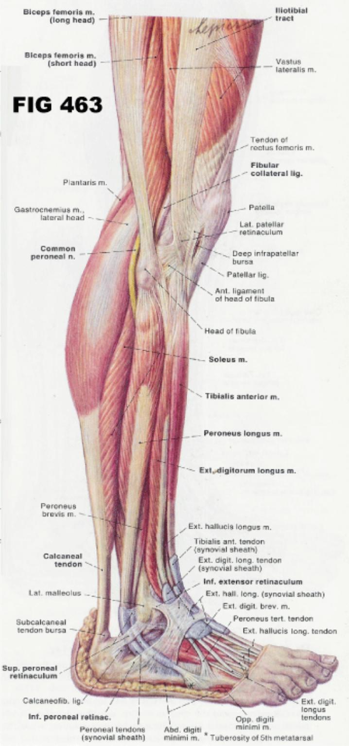
Fig 463
Superficial Muscles and Tendons of the Right Lower Thigh and Leg, Lateral View
1.5.2.22.3.1.1 The disposition of the anterior and lateral compartment muscles of the leg, and how their tendons, surrounded by tendon sheaths (in blue), enter the foot. Observe that the anterior compartment tendons enter the dorsum, while the lateral compartment tendons descend behind the lateral malleolus.
1.5.2.22.3.1.2
The superficial location of the head of the fibula and
its relationship to the common peroneal nerve.
1.5.2.22.3.1.3
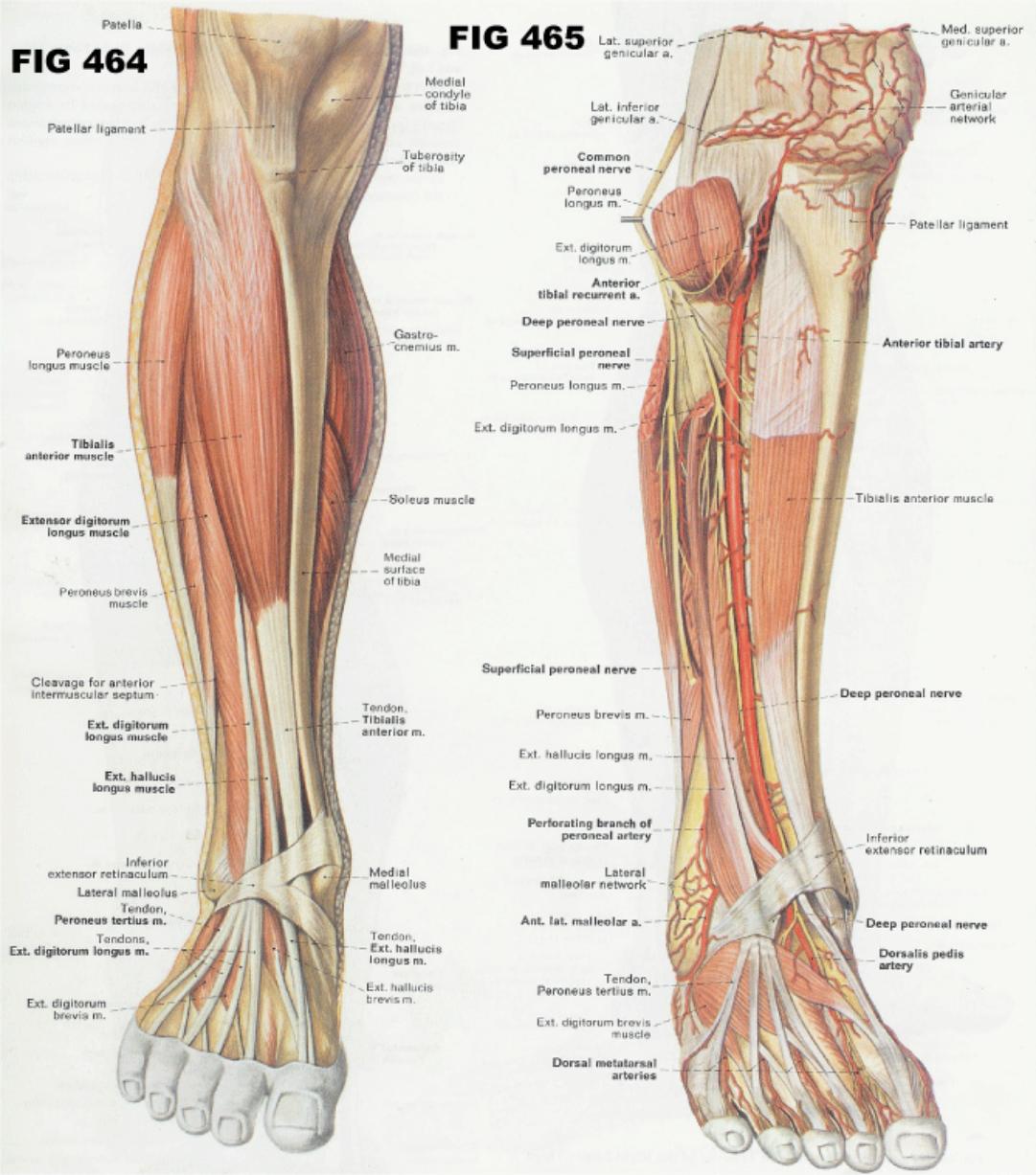
1.5.2.22.3.2
Fig
464 Muscles of the Anterior Compartment of the Leg
1.5.2.22.3.2.1 The four anterior compartment muscles are the tibialis anterior, extensor hallucis longus, extensor digitorum longus and peroneus tertius.
1.5.2.22.3.2.2
The tibialis anterior dorsally flexes and supinates the
foot. The other muscles extend the toes as well as dorsiflex the foot. Additionally,
the extensor hallucis longus assists in supination, while the extensor
digitorum longus and peroneus tertius are pronators.
1.5.2.22.3.3
Fig
465 Nerves and Arteries of the Anterior and Lateral Compartments of the Leg
1.5.2.22.3.3.1 As the common peroneal nerve courses laterally around the head of the fibula, it divides into the superficial and deep peroneal nerves which innervate the muscles of the lateral and anterior compartments.
1.5.2.22.3.3.2
The deep
peroneal nerve is joined by the anterior tibial artery which descends toward
the foot.
1.5.2.22.3.3.3
1.5.2.22.3.4
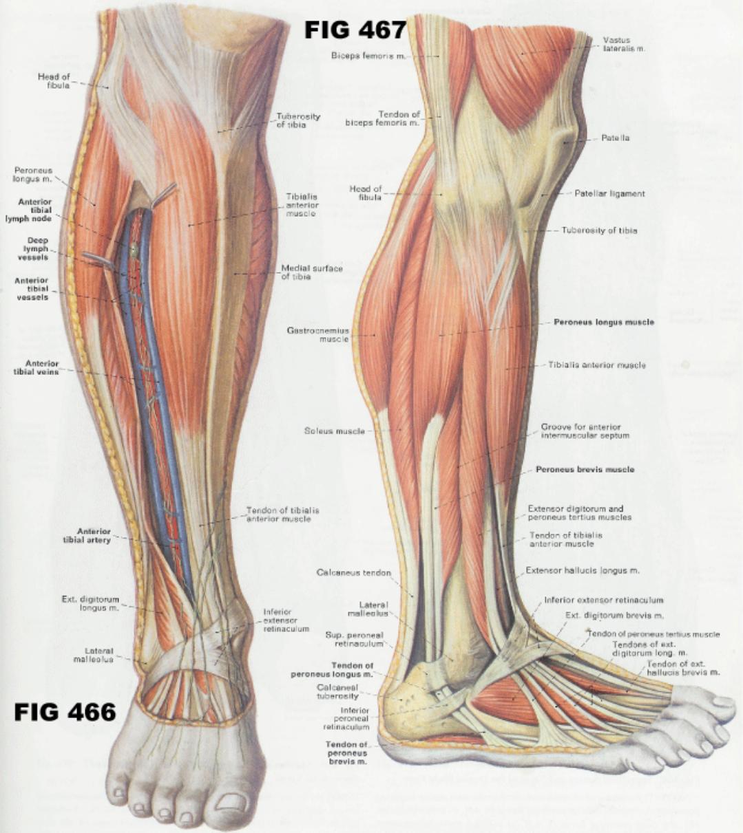
Fig
466 Deep Lymphatic Channels and Nodes of the anterior Leg
1.5.2.22.3.4.1
Lymphatic
channels from the dorsum of the foot course superiorly and collect along the
path of the more deeply situated anterior tibial vessels and nerve. At times a
lymph node can be found just ventral to the anterior tibial artery below the
knee.
1.5.2.22.3.5
Fig
467 Muscles of the Lateral Compartment of the Leg
1.5.2.22.3.5.1
The peroneus
longus and brevis occupy the lateral compartment of the leg. Their tendons
descend into the foot behind the lateral malleolus. The peroneus longus tendon
crosses the sole of the foot to insert on the base of the 1st metatarsal bone,
while the peroneus brevis inserts directly onto the 5th metatarsal bone.
1.5.2.22.3.5.2
1.5.2.22.4 Anterior Compartment
1.5.2.22.4.1 Extensor Digitorum Longus
1.5.2.22.4.1.1 This muscle dorsiflexes and everts the foot and extends the toes. Its origin is on the lateral condyle of the tibia, anterior surface of the fibula, and interosseous membrane. It inserts on the middle and distal phalanges of the four outer toes. It is supplied by the deep peroneal nerve with nerve roots of L4, L5 and S1.
1.5.2.22.4.2 Extensor Hallucis Longus
1.5.2.22.4.2.1 This muscle dorsiflexes and inverts the foot. Its origin is on the anterior surface of the fibula and interosseous membrane and it inserts on the distal phalanx of the great toe. It is supplied by the deep peroneal nerve with nerve roots of L4, L5 and S1.
1.5.2.22.4.3 Peroneus Tertius
1.5.2.22.4.3.1 This muscle dorsiflexes and everts the foot. Its origin is on the distal third of the fibula and interosseous membrane and it inserts on the fifth metatarsal. It is supplied by the deep peroneal nerve with nerve roots L4, L5 and S1.
1.5.2.22.4.4 Tibialis Anterior
1.5.2.22.4.4.1 This muscle dorsiflexes and inverts the foot. Its origin is on the lateral condyle and body of the fibia and interosseous membrane and it inserts on the first metatarsal and first (medial) cuneiform. It is supplied by the deep peroneal nerve with nerve roots of L4, L5 and S1.
1.5.2.22.5 Lateral Peroneal Compartment
1.5.2.22.5.1 Peroneus Brevis
1.5.2.22.5.1.1
This muscle plantar flexes and everts the foot. Its
origin is on the body of the fibula and it inserts on the base of the fifth
metatarsal. It is supplied by the superficial peroneal nerve with nerve roots
of L4, L5, S1, & S2.
1.5.2.22.5.2 Peroneus Longus
1.5.2.22.5.2.1 This strap muscle along with the Tibialis posterior help support the medial transverse arch of the foot. This muscle plantar flexes and everts the foot. Its origin is on the head and body of the fibula and lateral condyle of the tibia and it inserts at the first metatarsal and first cuneiform. It is supplied by the superficial peroneal nerve with nerve roots of L4, L5, S1, & S2.
1.5.2.22.6 Posterior Superficial Compartment
1.5.2.22.6.1 Gastrocnemius
1.5.2.22.6.1.1 This muscle plantar flexes the foot and flexes the leg. Its origin is on the lateral and medial condyles of the femur and capsule of the knee and it inserts on the calcaneus by way of the calcaneal (Achilles) tendon. It is supplied by the tibial nerve with nerve roots of S1, & S2.
1.5.2.22.6.2 Plantaris
1.5.2.22.6.2.1
This muscle plantar flexes the foot. Its origin is on
the femur above the lateral condyle and it inserts onto the calcaneus by way of
the calcaneal (Achilles) tendon. It is supplied by the tibial nerve and its
nerve roots are L4, L5, S1, & S2.
1.5.2.22.6.2.2
1.5.2.22.6.3 Soleus
1.5.2.22.6.3.1 This muscle attaches to the upper portion of the lower leg and to the Achilles tendon. During the stance phase of gate, it checks the forward motion of the lower leg and plantar flexes the foot during the gate phase of push-off. While seated this one joint muscle is a strong plantar flexor while the gastrocnemius is mechanically disadvantaged due to its lengthened position. It is supplied by the tibial nerve with nerve roots of L5, S1, & S2.
1.5.2.22.7 Posterior Deep Compartment
1.5.2.22.7.1 Flexor Digitorum Longus
1.5.2.22.7.1.1
This muscle plantar flexes and inverts the foot and
flexes the toes. Is origin is the posterior surface of the tibia and it inserts
onto the distal phalanges of the four outer toes. It is supplied by the tibial
nerve with nerve roots of L5, S1, S2, S3.
1.5.2.22.7.1.2
1.5.2.22.7.2 Flexor Hallucis Longus
1.5.2.22.7.2.1 This muscle plantar flexes and inverts the foot and flexes the big toe. Its origin is on the lower two-thirds of the fibula and it inserts onto the distal phalanx of the big toe. It is supplied by the tibial nerve and its nerve roots are S2-3.
1.5.2.22.7.3 Popliteus
1.5.2.22.7.3.1 This muscle flexes and medially rotates the leg. Its origin is on the lateral condyle of the femur with insertion on the proximal tibia. It is supplied by the tibial nerve and its nerve roots are L4, L5, & S1..
1.5.2.22.7.4 Tibialis Posterior
1.5.2.22.7.4.1 This strap muscle along with the Peroneus longus and brevis help support the medial transverse arch of the foot. The Tibialis posterior is the deepest posterior lower leg muscle attaching to the Interosseous Membrane (fibrous connective tissue which binds the tibia and fibula) and to both the tibia and fibula covering a major portion of the lower leg. This muscle also attaches to many of the bones that wedge together in forming the Roman style arch of the foot including the bases of the middle three metatarsal bones. This muscle plantar flexes and inverts the foot. Its origin is on the tibia, fibula, and interosseous membrane and it inserts onto the second, third, and fourth metatarsals; navicular, all three cuneiforms, and cuboid. It is supplied by the tibial nerve and its nerve roots are L4-5.
1.5.2.23 Foot-Intrinsic *
Back Table of Contents References
1.5.2.23.1
Introduction
1.5.2.23.1.1
The
intrinsic muscles of the foot are similar to the hand muscles, which are
specialized for intricate and precise movements where as the foot muscles are
specialized for support and locomotion. The deep facia of the foot forms the
plantar aponeurosis (fascia) that attaches to the calcaneus and the phalanges
providing longitudinal arch support. The intrinsic foot muscles are divided
into two groups; Dorsal, which includes only two muscles (see below) and the plantar muscles, which
include several layers as, outlined below.
1.5.2.23.2
Gray’s
Anatomy
1.5.2.23.2.1
Intrinsic Foot Muscles
1.5.2.23.3 Dorsal Muscles
1.5.2.23.3.1 Extensor Digitorum Brevis
1.5.2.23.3.1.1 This muscle extends the first through fourth toes. Its origin is on the dorsal aspect of the calcaneus and it inserts on the tendon of the extensor Digitorum longus and proximal phalanx of the great toe. It is supplied by the Deep peroneal nerve with nerve roots of S1 and S2.
1.5.2.23.3.2 Extensor Hallucis Brevis
1.5.2.23.3.2.1 This muscle extends the proximal phalanx of the Hallux. Its origin is on the dorsal aspect of the calcaneus and it inserts on the dorsal surface of the base of proximal phalanx of Hallux. It is supplied by the deep peroneal nerve with nerve roots of S1 and S2.
1.5.2.23.4 Plantar Muscles
1.5.2.23.4.1 Plantar First Superficial Layer
1.5.2.23.4.1.1 Abductor Digiti Minimi (Foot) B1E1
1.5.2.23.4.1.1.1 The action of this muscle is to abduct the fifth toe away from the fourth toe. This muscle has its origin on the calcaneus and it inserts onto the little toe. It is supplied by the lateral plantar nerve with nerve roots of S2 and S3.
1.5.2.23.4.1.2 Abductor Hallucis B3E3
1.5.2.23.4.1.2.1 The action of this muscle is to abduct the big toe from the mid line of the foot. This muscle has its origin on the calcaneus and it inserts onto the big toe. It is supplied by the medial plantar nerve with nerve roots of L4, L5, S1, S2, & S3.
1.5.2.23.4.1.3 Flexor Digitorum Brevis
1.5.2.23.4.1.3.1
The action of this muscle is to flex the second through
fifth toes. This muscle has its origin on the calcaneus and plantar aponeurosis
and it inserts onto the middle phalanx of the second through fifth toes. It is
supplied by the medial plantar nerve with nerve roots of L4,
L5, S1, S2, & S3.
1.5.2.23.4.1.4 Sectional Questions
1.5.2.23.4.1.4.1 Questions
1.5.2.23.4.2 Plantar Second Layer
1.5.2.23.4.2.1.1 This muscle extends the second through fifth toes. Its origin is on the tendons of the flexor Digitorum longus and it inserts onto the tendons of the extensor Digitorum longus. It is supplied by the medial and lateral plantar nerves with nerve roots of L4, L5, S1, S2, & S3.
1.5.2.23.4.2.2 Quadratus Plantae
1.5.2.23.4.2.2.1 This muscle flexes the second through fifth toes. Its origin is on the calcaneus and it inserts onto the tendons of the flexor Digitorum longus. It is supplied by the lateral plantar nerve with nerve roots of S2 and S3.
1.5.2.23.4.3 Plantar Third Layer
1.5.2.23.4.3.1 Adductor Hallucis B7E7
1.5.2.23.4.3.1.1 The adductor hallucis adducts the big toe towards the 2nd toe and Flexes the big toe towards plantar surface. The oblique head attaches (origin) to the bases of the 2nd, 3rd and 4th metatarsals. The transverse head attaches (origin) to the Plantar Metatarsophalangeal ligaments of the 3rd, 4th and 5th toes. Both heads insert into the lateral side of base of proximal phalanx of big toe. This muscle is supplied by the lateral plantar nerve and its nerve roots are S2 and S3.
1.5.2.23.4.3.2 Flexor Digiti Minimi Brevis
1.5.2.23.4.3.2.1 This muscle flexes the small toe. Its origin is on the fifth metatarsal and it inserts on the proximal phalanx of the small toe. It is supplied by the lateral plantar nerve and its roots are S2 and S3.
1.5.2.23.4.3.3 Flexor Hallucis Brevis
1.5.2.23.4.3.3.1 This muscle flexes the great toe. Its origin is on the cuboid and third (lateral) cuneiform with insertion onto the proximal phalanx of the great toe. It is supplied by the medial plantar nerve with nerve roots of L4, L5, S1, S2, & S3.
1.5.2.23.4.4 Plantar Fourth Deep Layer
1.5.2.23.4.4.1 Dorsal Interossei
1.5.2.23.4.4.1.1 This muscle abducts the toes and flexes the proximal phalanges. Its origin is on the adjacent side of the metatarsals and it inserts onto the proximal phalanges, both sides of the second toe, and the lateral side of the third and fourth toes. It is supplied by the lateral plantar nerve and its nerve roots are S2 and S3.
1.5.2.23.4.4.2 Plantar Interossei
1.5.2.23.4.4.2.1 This muscle adducts the third, fourth, and fifth toes and flexes the proximal phalanges. Its origin is on the third, fourth and fifth metatarsals and it inserts onto the proximal phalanges of the same toes. It is supplied by the lateral plantar nerve with nerve roots of S2 and S3.
1.5.3
Individual
Muscles
Back Table
of Contents References